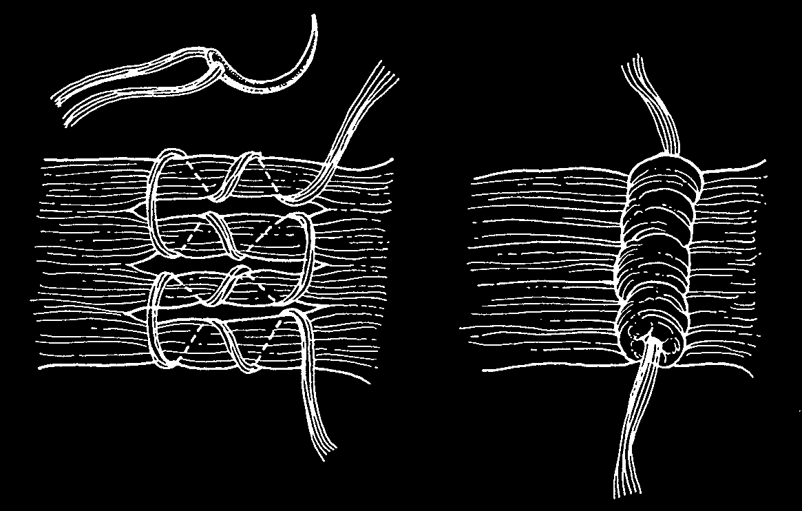
Figure 14
An ingenious technique, the O’Connor cinch, produced minimal shortening of a rectus muscle. A large, dull needle was used to weave a multiple-strand suture through slips of the tendon or muscle. The greater the number of threads, producing a bulkier suture and a greater diameter of thread, the more shortening of the muscle is produced.
A
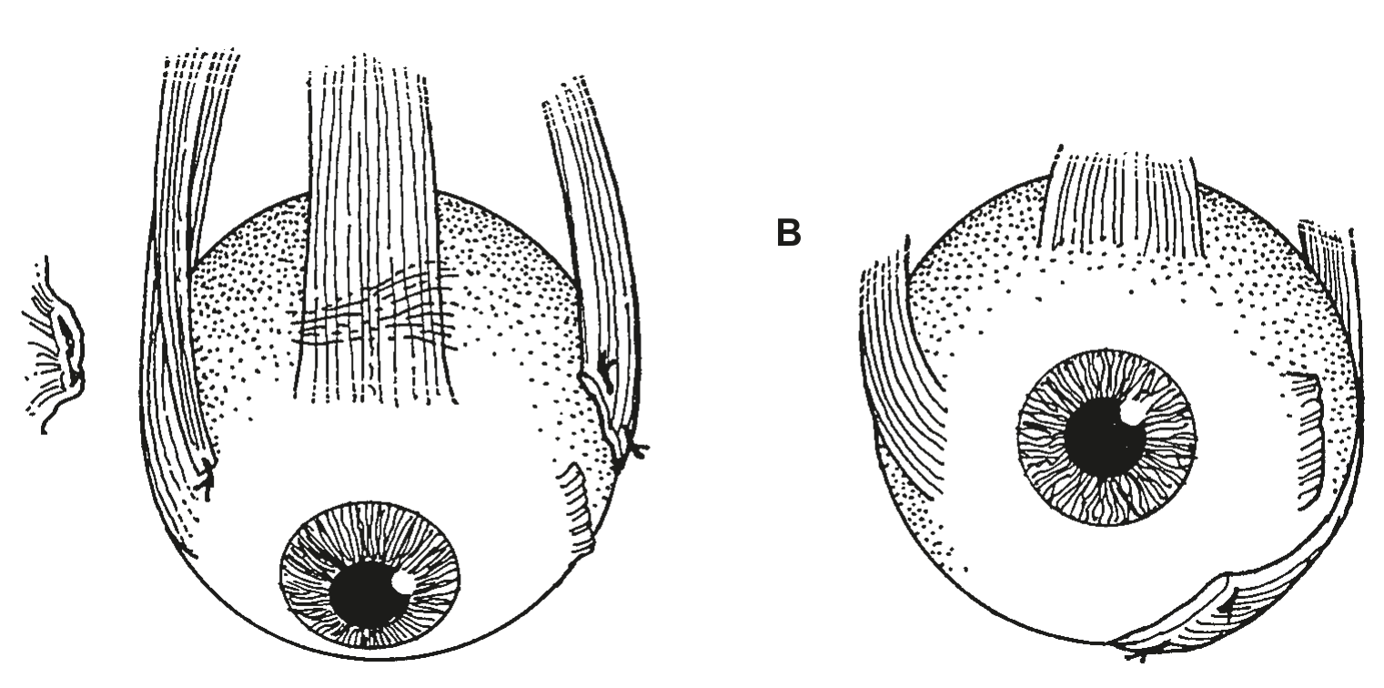
Figure 15
B
A Peter suggested fracturing the trochlea and then shifting and attaching the superior oblique muscle to a point near the medial rectus insertion. This procedure was combined with lateral rectus tenotomy or recession for treatment of third cranial nerve palsy.
B The lateral rectus was shifted for treatment of congenital absence of the inferior rectus.
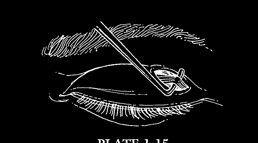
Figure 16
A transcutaneous approach to the superior oblique tendon afforded exposure for tenectomy or tucking. Skin incisions were commonly used for exposure of both superior and inferior oblique muscles in the early twentieth century.
A1
A2
B
C
D
E
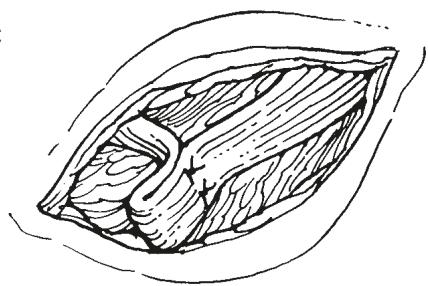
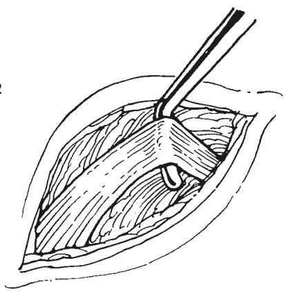
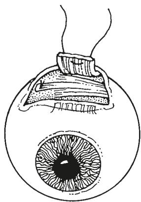
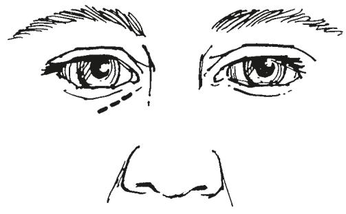
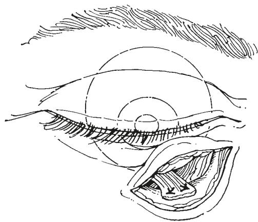
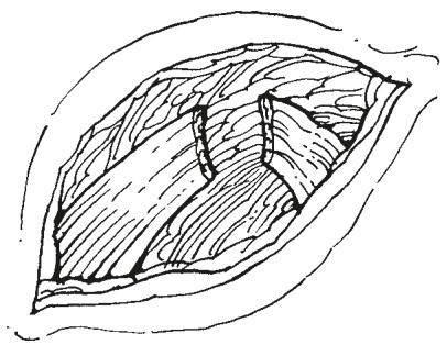
Figure 17
A1 Wheeler exposed the inferior oblique muscle nasal to the inferior rectus through a skin incision.
A2 The inferior oblique muscle was then engaged with a muscle hook.
B From that exposure the muscle could be weakened by myotomy.
C The muscle also could be shortened by a tuck.
D A curious inferior oblique ‘strengthening’ procedure performed by Wheeler was disinsertion of the muscle followed by reinsertion of the cut end into the inferior orbital rim periosteum.
E Wheeler strengthened the superior oblique muscle by advancing the central portion of the tendon. He recognized that this procedure produced decreased elevation in adduction. In addition, he recognized that shifting the tendon anteriorly produced intorsion and that posterior fibers influenced depression of the globe, a concept later credited to Harada and Ito (Jpn J Ophthalmol 8:88, 1964).