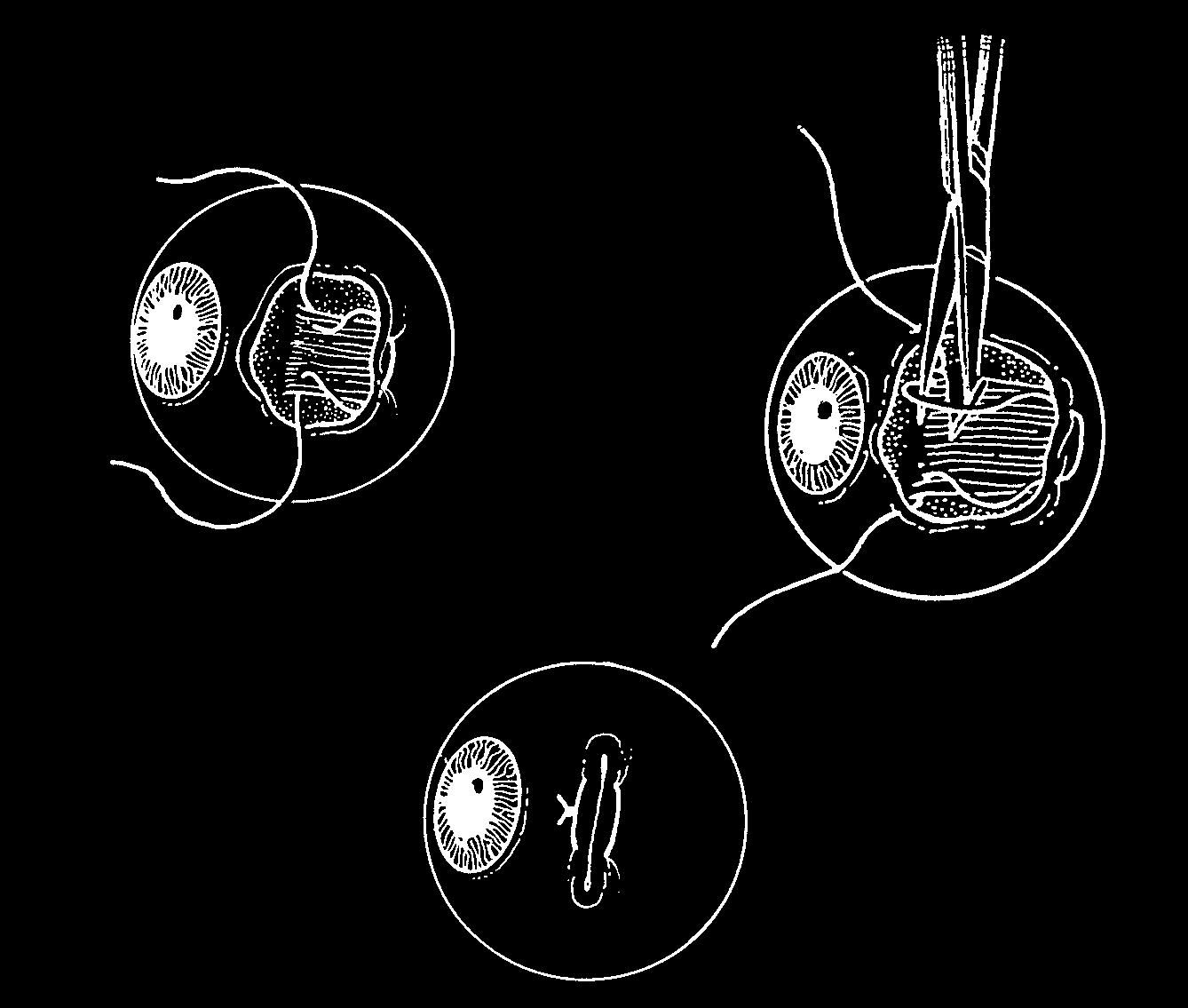
Figure 7
Most early recession procedures involved sutures entering and exiting the conjunctiva for added security and to facilitate removal of silk sutures. Tenon’s capsule and the muscle capsule were incorporated in recessions and resections. The muscle is cut between bites of the preplaced suture to avoid ‘loss’ of the disconnected proximal muscle end. Externally placed buttons or bolsters added additional support to the recessed muscle, facilitated suture adjustment, and made suture removal easier.
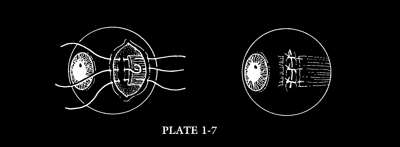
Figure 8
Jameson’s technique for recession included several sutures anchoring the muscle to the sclera. The external sutures could be removed and loosened or tightened postoperatively if a significant overcorrection or undercorrection was produced.

Figure 9
A Early needles were round; had eyes for threading the suture; and in most cases, had a wire diameter greater than the thickness of the sclera. These factors made the ophthalmic needle a ‘formidable weapon.’
B Jameson admonished surgeons to keep the needle in view while passing it through the sclera, to avoid entering the globe and creating what he described as panophthalmia.
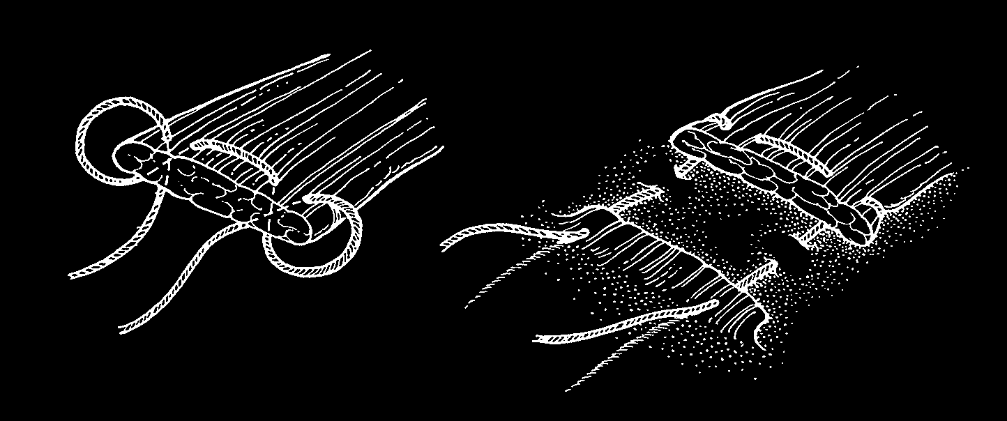
Figure 10
Peter’s whip stitch for securing the cut end of the muscle during recession of a rectus muscle is a sound technique that is still used today.
A
B
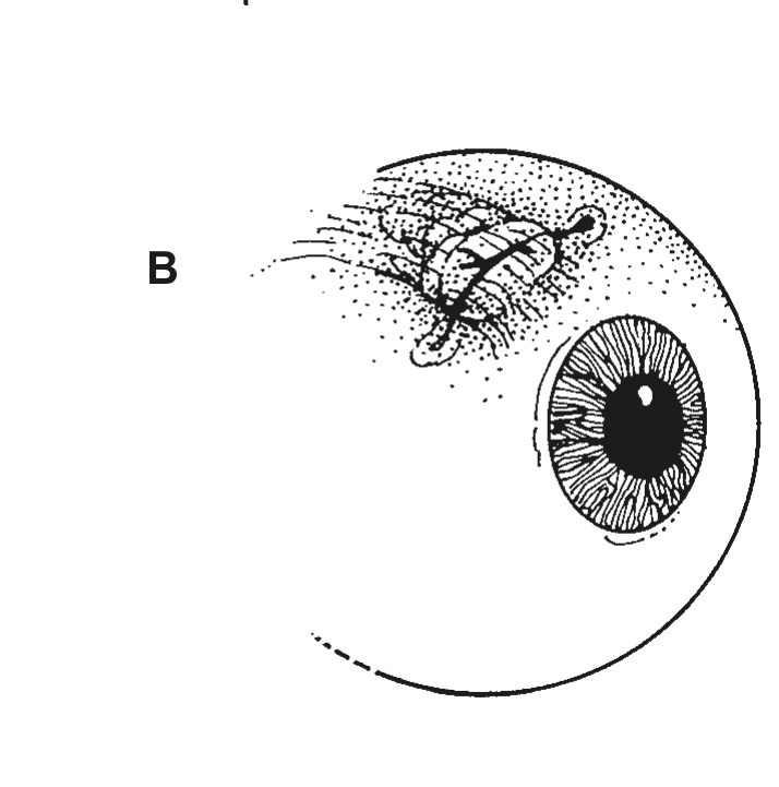
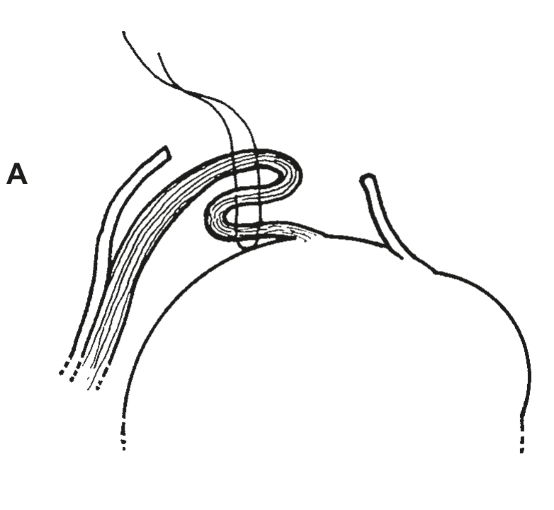
Figure 11
A A simple tuck for muscle-tendon shortening (‘strengthening’) avoided the need for placing a needle through the sclera and produced a shortened muscle without removing a piece.
B When performed near the limbus, tucking tended to produce unsightly bulk that could be cosmetically disfiguring.
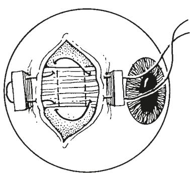
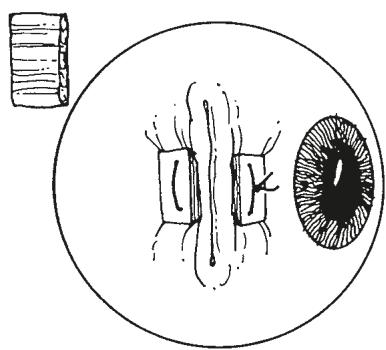
Figure 12
Pragnen used gold buttons as bolsters to eliminate ‘cheese wiring’ of the externalized suture when carrying out resection of a rectus muscle. This technique allowed postoperative adjustment of the length of the muscle.
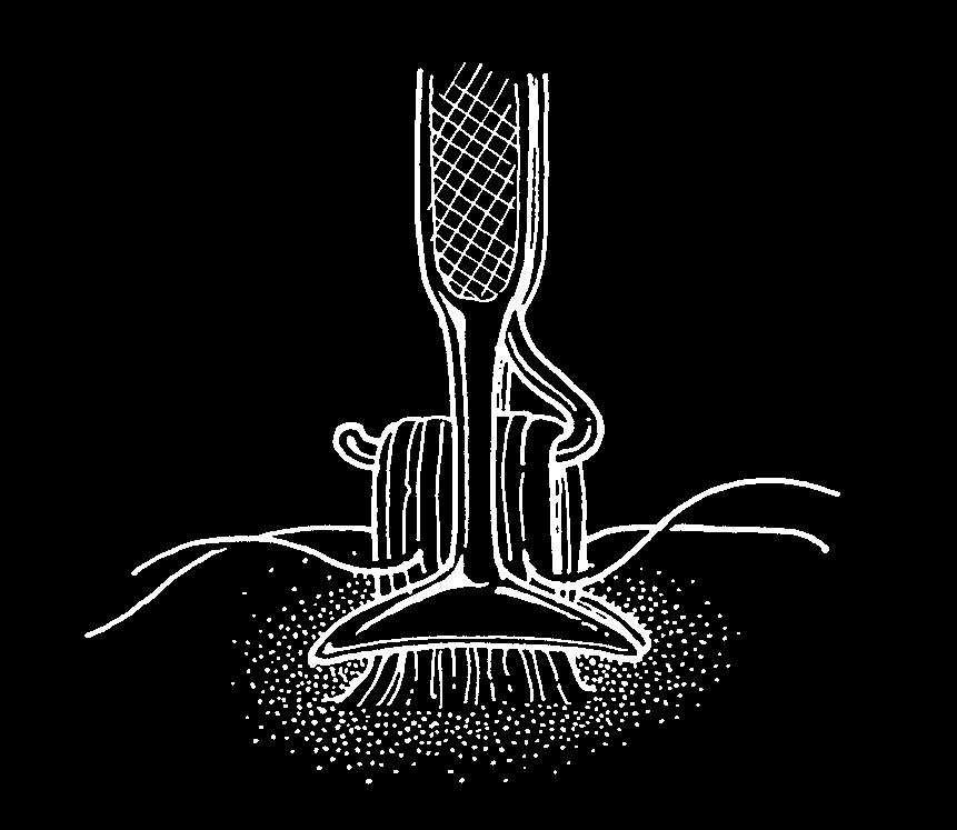
Figure 13
Peter’s modification of the Bishop tucking instrument was one of several elegant instruments that were designed to facilitate the tucking procedure. Tucking of the extraocular muscles was an early favorite muscle shortening ‘strengthening’ procedure.