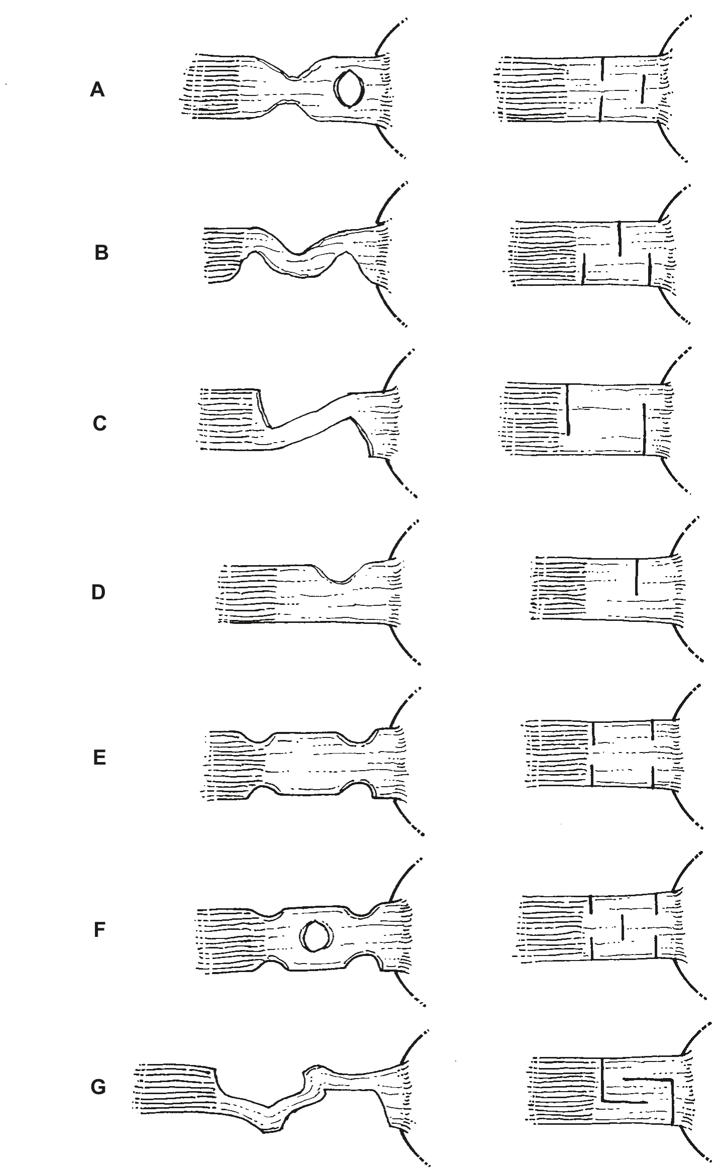
Figure 4
A
B
C
D
E
F
G
A wide variety of tendon lengthening procedures provided a safeguard against overcorrection and could also be carried out without sutures, The simple Z tenotomy (Blaskovics, 1912) is still used today for weakening an already maximally recessed muscle (Chapter 4).
A O’Connor
E Abadie
B Bishop-Harmon
F Verhoeff
C Blaskovics
G Terrien
D von Graefe
Some modern surgeons are reporting satisfactory clinical results from tenotomy which does not cut across all fibers, but more or less nicks the muscle. Biglan reports success treating small vertical deviations by doing incomplete tenotomy of the superior rectus.

Figure 5
The use of sutures added to the complexity and safety, if not the effectiveness, of tenotomy. Again, the principle was that a lengthened tendon-muscle would result in reduced muscle pull and therefore produce a change in alignment, shifting the globe ‘away’ from the weakened muscle.
A
B
C
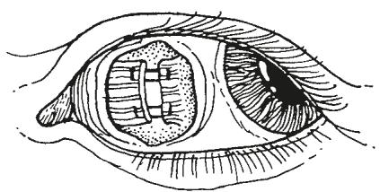

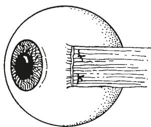
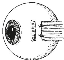
Figure 6
A Sutures were also used to reapproximate myotomized muscles to avoid producing more than the intended result. The sutures were not placed in the sclera. They were placed only in the cut ends of the muscle and tendon. The initial use of sutures apparently was for overcorrected and not primary cases.
B Prince (1887) was credited with describing recession with the tendon actually sutured to the sclera. This procedure was the forerunner of more accurate and reproducible surgery.
C Worth (1904) described resection using sutures. This muscle shortening was accomplished with or without recession of the antagonist muscle.
Late in the nineteenth century, measured recession with reattachment of the muscle to the sclera was carried out. Tendon and muscle tucking or plication; muscle advancement procedures; and, later, resection and advancement procedures were also performed (Figure 6).
Surgery of the oblique muscles began when inferior oblique weakening was carried out to treat myopia and asthenopia, not surprisingly without success. The superior oblique muscle was considered by some noli me tangere, loosely translated today as ‘off limits,’ in the early twentieth century. However, all types of oblique surgery had been described by the middle part of the twentieth century. Fink, in 1951, said “up to a few years ago, all corrective surgical measures for the obliques were not considered feasible because of anatomical difficulty.”*
Along with oblique muscle surgery and vertical rectus muscle recession and resection, extraocular muscle transfer procedures were used for strabismus caused by muscle paralysis. Although, at first, the improved alignment after eye muscle transfer procedures was thought to be due to postoperative neurologic reorientation, it became apparent from electromyographic studies that the effect of an extraocular muscle transfer procedure was mediated through mechanical factors.
Technical advances in strabismus surgery have occurred along with, and in some ways because of, advances in anesthesia. A glass of wine and a good lunch followed by a head lock and a quick surgeon were early ‘anesthesia.’ This was replaced by pontocaine, cocaine, and ether for general anesthesia, and later, a wide variety of modern agents were used. Supporting modern anesthetic agents are the extensive and detailed monitoring devices that provide instantaneous, real-time recordings of temperature, respiration, electrocardiographic data, and oxygen saturation.
Highlights of the history of strabismus surgery from its earliest beginnings in 1739 to the present are depicted in the following drawings. They describe a wide variety of manipulations that surgeons employed to straighten the eyes by altering the state of the extraocular muscle (Figures 7-24). The statement has been made, “Those who do not learn from history are destined to repeat it.” Thus, some surgical procedures have a tendency to be rediscovered every generation or so by surgeons who may have overlooked an earlier description. Nonetheless, strabismus surgery has followed a steady progression, with improvement through innovations in the surgical technique, combined with better instruments, more reliable sutures, safer anesthesia, and a better appreciation on the part of the surgeon of neural, sensory, and mechanical factors.
*Fink WH: Surgery of the oblique muscles of the eye, St Louis, 1951, The CV Mosby Co.