Section 1
Chapter 1: History of strabismus surgery
Chapter 2: Surgical anatomy
Chapter 3: Parasurgical procedures and preparation
1
History of strabismus surgery
Strabismus, recognized from the earliest times because the eyes are readily observable, has changed little in form throughout the years. However, its causes are now well understood, and its treatment undergoes regular revision and refinement. The condition, at first, was considered to be a visitation of an evil spirit and incurable. The earliest physicians suggested treatment with the only available methods-- potions, purification, and diet. In the seventh century, masks designed to redirect the visual axes were described. Curiously, variations of this type of treatment are still advocated by some today in the form of sector occlusion, despite 13 centuries of failure!
The practice of strabismus surgery began inauspiciously in 1739 with the efforts of John Taylor (Figure 1). He is reported to have had “...a considerable amount of sense...,” according to Stewart Duke- Elder, who said Taylor undoubtedly recognized that strabismus was a muscle abnormality that could be treated by dividing the extraocular muscles.* However, Taylor’s surgery was not successful. On the contrary, Taylor is said to have been a showman who may have only snipped the conjunctiva, patched one eye, and left town before the results could be assessed. By patching the unoperated eye and having the operated eye take up fixation, Taylor’s procedure would have given the appearance of being successful since the operated eye would appear to have been ‘straightened.’ John Taylor’s position in history appears to be one of ridicule rather than honor.
After Taylor, several surgeons contributed to the body of knowledge that led to successful, scientifically founded strabismus surgery. In 1752, Eschenbach made the comment “each oculist dreams from time to time that it may be possible to dissect completely or partially the offending rectus muscle”.
In 1816 Dulspech, an orthopedic surgeon, performed tenotomy of the Achilles tendon as treatment for club foot with a technique not too different from strabismus surgery. Gibson experimented with extraocular muscle transection for treatment of esotropia in 1818 but did not report this until 1841. White, in 1827, made similar attempts without success. Stromeyer performed extraocular muscle tenectomy on a cadaver in 1838, and a year later, Dieffenbach performed successful myotomy of the medial rectus on a 7-year-old boy with esotropia. His priority was contested by Cunier, but the honor has been granted to Dieffenbach. The year 1839 is generally considered to be the beginning of modern strabismus surgery.
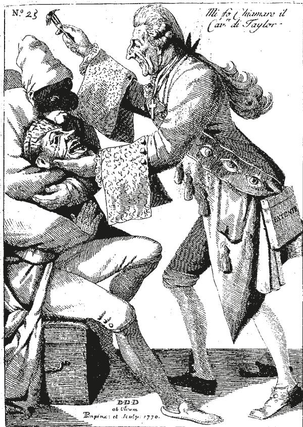
Figure 1
Abundant showmanship and fast carriage rather than surgical skills were outstanding attributes of the first strabismus ‘surgeon’ John Taylor (1703-1772), an itinerant healer who is depicted in this eighteenth century drawing.
*Duke-Elder S and Wybar K: System of ophthalmology, Vol 6, Ocular motility and strabismus, St Louis, 1973, The CV Mosby Co. †von Noorden GK (ed): History of Strabismology, Belgium 2002, J. P. Wayenborgh
In the nearly century and a half since its beginning with Dieffenbach, strabismus surgery has undergone many refinements, but the basic principles have changed little. The orthopedic principles that were adhered to at the beginning of strabismus surgery remain as the primary guiding factors today. To these have been added new knowledge of the central nervous system influences and psycho-physical factors related to strabismus. Understanding of neural mechanisms combined with knowledge and appreciation of both the dynamic muscle forces and the physiologic and pathologic restrictive aspects of ocular motility make strabismus surgery a science. Improved diagnostic techniques, such as passive duction testing, interpretation of differential intraocular pressure, differential saccadic velocity measurement, electrooculogram testing of nystagmus, and generated muscle force testing, have enabled more accurate diagnosis leading to more accurate surgery.
Improved imaging techniques, especially those using magnetic resonance imaging (MRI), have made possible the noninvasive morphologic study of extraocular muscles. Technical improvements in anesthesia, sutures and needles, implanted materials, antibiotics, corticosteroids, and, most recently, injectable toxins have broadened the scope of strabismus surgery. Recent studies of the origins of the most common strabismus, essential infantile or congenital esotropia, have promoted earlier surgery. Strabismus surgery, frequently done as an office procedure in its early years, is now once again done on an outpatient basis.
Strabismus surgery began with weakening procedures of the medial rectus. These procedures were first performed by transecting the muscle and often produced disastrous results (Figure 2). Shifting surgery from the belly of the muscle to the tendon reduced the number of overcorrections (Figure 3). Tendon weakening or lengthening, which avoided cutting across the full width of the tendon, was subsequently carried out using a variety of ingenious techniques. The earliest of these were performed without placing a needle and suture through the tendon. Instead, one or more incisions were made in the muscle or tendon, usually at right angles to the long axis of the muscle and crossing the midline. Later, tendon lengthening procedures used sutures. (Figure 4 & 5).

Figure 2
Taylor may have only snipped the conjunctiva, patched the eye, and then left town. He is said to have operated twice on Bach, the musician of Leipzig, who later became blind. The antics of Taylor may have been responsible for a 100- year delay in the establishment of strabismus surgery as part of the surgeon’s armamentarium.
A
B

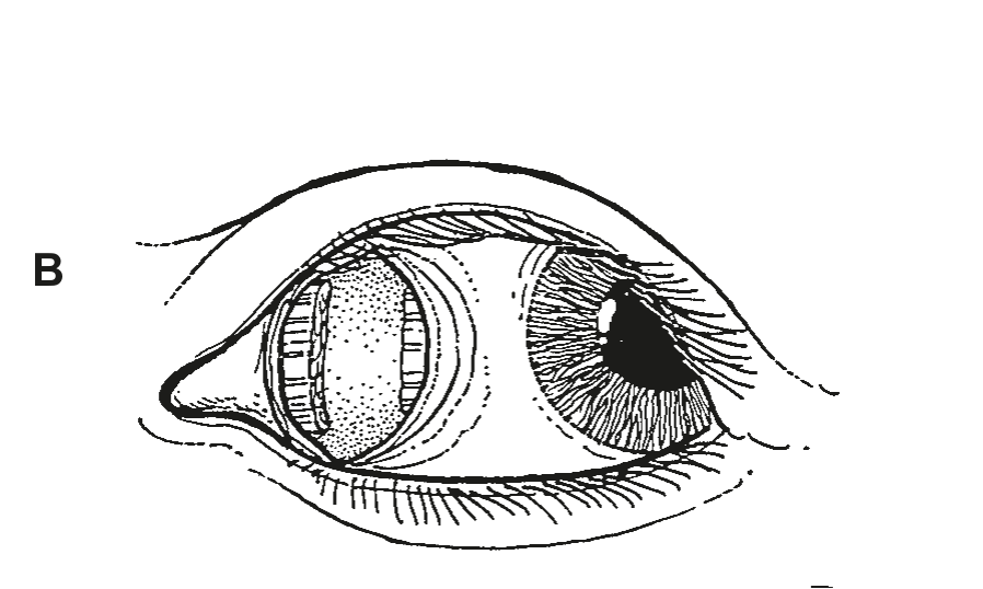
Figure 3
A Myectomy of the medial rectus for esotropia, as performed by Dieffenbach in 1839, often produced a large exotropia and unsightly medial displacement of the caruncle. The so-called cure may have been only a slight improvement over the malady.
B Simple tenotomy carried out nearer the insertion reduced the number and severity of overcorrections and lessened caruncle displacement.