We find the technique of rectus muscle palpation with a blunt instrument to be highly useful when identifying the borders of the rectus muscles intraoperatively, and often find the palpation technique more valuable than visual inspection. To perform this technique, the eye is rotated with fixation forceps or bridle sutures into the desired position for surgery (>Fig. 1.16). A blunt instrument, such as a muscle hook, is placed on the conjunctiva between two adjacent rectus muscles approximately 10 mm posterior to the corneal limbus. While applying gentle posterior pressure on the globe with the muscle hook, the hook is directed toward the rectus muscle. The border of the muscle can be easily palpated in this manner and readily visualized as the hook makes contact with the border of the muscle, assuring the surgeon that the eye has been properly positioned for surgery and helping to facilitate accurate placement of the conjunctival incision.
1.7 Innervation of the Extraocular Muscles
The third cranial nerve (oculomotor nerve) is the most complex of the cranial nerves supplying innervation to the extraocular muscles. It provides the innervation to four of the six extraocular muscles and to the levator palpebrae superioris muscle of the upper eyelid. The inferior division of the third nerve supplies the medial and inferior rectus muscles as well as the inferior oblique muscle. The superior division supplies the superior rectus muscle and levator palpebrae superioris muscle of the upper eyelid. Motor branches of the third nerve enter the medial and inferior rectus muscles at approximately the junction between the posterior one-third and anterior two-thirds of the muscle from the internal or global surface of the muscle. The neurovascular bundle supplying the inferior oblique muscle enters the muscle from its posterior surface near the lateral border of the inferior rectus muscle [20].
The superior oblique muscle is innervated by the fourth cranial nerve (trochlear nerve). It is the only extraocular muscle that receives its innervation from the external or orbital surface of the muscle. The nerve passes superiorly from the medial side of the superior oblique muscle to the orbital side of the muscle prior to entering the muscle as several small branches.
The lateral rectus muscle is innervated by the sixth cranial nerve (abducens nerve). The muscle is innervated from the internal or bulbar surface of the muscle near the junction of the anterior two-thirds and posterior one-third of the muscle.
1.8 Blood Supply to Extraocular Muscles
Each of the extraocular muscles receives its blood supply from the medial and lateral muscular branches of the ophthalmic artery. The medial branch supplies the inferior and medial rectus muscles as well as the inferior oblique muscle, while the lateral branch supplies the lateral and superior rectus muscles, the superior oblique muscle, and the levator muscle of the upper eyelid. The inferior rectus muscle and inferior oblique muscle also receive a small contribution of blood supply from other sources.
The arteries of the four rectus muscles enter the muscles on their global surfaces at approximately the junction between the anterior two-thirds and posterior third of the muscle. They course anteriorly, emerging onto the orbital surface of the muscle/tendon approximately 10–12 mm from the insertions of the tendons into the sclera, where they are known as the anterior ciliary arteries. Each rectus muscle characteristically has two anterior ciliary arteries, except for the lateral rectus muscles, which characteristically have only one (>Fig. 1.17). The course of the anterior ciliary arteries along the muscles and their tendons is highly variable and this variation is important to recognize when planning surgery on patients who are at risk for anterior segment ischemia (Chap. 20). The anterior ciliary vessels course forward to the episclera, where they supply branches to the sclera, limbus and to the conjunctiva. They enter the sclera near the limbus where they ultimately anastomose with the long ciliary arteries to form the major arterial circle of the iris. Veins corresponding to the muscle’s arteries drain into the superior and inferior orbital veins.
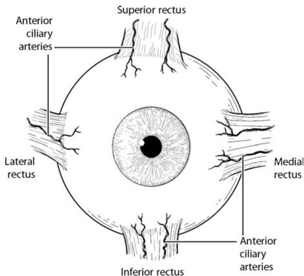
Fig. 1.17. The anterior ciliary arteries. Except for the lateral rectus muscle, each rectus muscle contains two anterior ciliary arteries
1.9 Surgically Important Anatomy of Individual Extraocular Muscles
This section will review key elements of the surgical anatomy of the individual extraocular muscles and surrounding tissues that the strabismus surgeon is likely to encounter frequently during standard strabismus surgery. Information about the basic anatomy, function, and structure of each extraocular muscle and its supporting fascia is found elsewhere in this and subsequent chapters.
1.9.1 Medial Rectus Muscle
The medial rectus muscle insertion is typically closer to the corneal limbus than that of the other rectus muscles. According to Apt [18] in the adult eye, the mean insertion distance from the center of the insertion to the anterior limbus is 5.3 mm (>Fig. 1.12). There is a wide range in variation of this distance [18] and this distance is smaller in a child. As a general rule of thumb, the surgeon should expect to find the insertion 1 mm closer to the limbus than the values shown in Table 1.1 for a child between the ages of 6 and 12 months. Helveston believes that variation in the insertion of the medial rectus muscle tendon relative to the limbus is large and common, suggesting a range of 3.5–6 mm from the corneal limbus [21]. He believes that the large variation in distance of the insertion of the medial rectus muscle relative to the corneal limbus makes the muscle insertion a poor landmark for measuring during recession surgery on the medial rectus muscle and prefers to utilize the limbus as the landmark, rather than the muscle insertion.
The medial rectus muscle is the only extraocular muscle that does not have a direct attachment to one of the other extraocular muscles. This increases the likelihood that the medial rectus muscle will retract posteriorly into the orbit if extensive dissection of the medial rectus muscle fascial system is carried out and the surgeon loses control of the muscle after it has been detached from the sclera. The surgeon is not likely to engage other vascular or muscular structures when hooking the medial rectus muscle.
The sclera posterior to the rectus muscle insertions is thinner than the sclera anterior to the rectus muscle insertions. This is particularly true of the region posterior to the medial rectus muscle insertion. Small recessions may require fixation of the muscle to this very thin portion of the sclera, where the risk of scleral perforation is probably higher, and may warrant consideration of a muscle hang-back procedure from the thicker sclera at the insertion in some situations. The underlying uvea can sometimes be seen through the thin sclera posterior to the media rectus muscle insertion and can occasionally be a cause of significant concern to patients postoperatively (Chap. 19). The conjunctival anatomy of the medial aspect of the eye is more complex than the conjunctiva encountered when performing surgery on the other rectus muscles (>Fig. 1.18). Care must be taken during the creation of conjunctival incisions at the start of surgery and during closure of conjunctival incisions at the conclusion of surgery to avoid disruption of the key anatomical landmarks of the medial conjunctiva, which can result in very significant cosmetic and functional complications (Chap. 19).