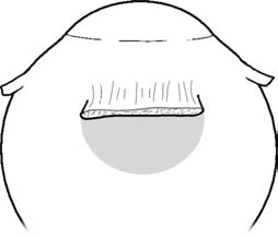
Fig. 1.7. Thin sclera posterior to the rectus muscle insertions, highlighted area
1.4 Fascial System
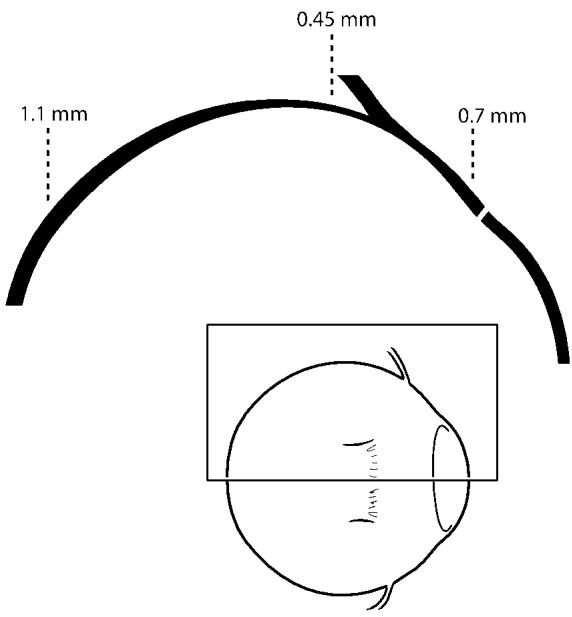
Fig. 1.8. Thickness of the sclera varies depending on its position around the globe
1.4 Fascial System
1.4.1 Tenon’s Fascia
The globe is suspended within the bony orbit by a fascial system, the bulk of which is represented by Tenon’s capsule. Tenon’s capsule is a condensation of fibrous tissue that covers the globe from the entrance of the optic nerve into the posterior aspect of the globe extending to within 1 mm of the corneal limbus, where it becomes fused with the overlying conjunctiva. Tenon’s capsule is thick and readily manipulated surgically in young patients, but becomes thin and fragile in older patients. Potential spaces exist both deep and external to Tenon’s capsule, known as the episcleral (sub-Tenon’s space) and the subconjunctival spaces, respectively. We will generally utilize the term episcleral space in this textbook to refer to the potential space deep to Tenon’s capsule. These spaces are important during strabismus surgery as they must be entered in order to gain access to the extraocular muscles. The anterior aspect of Tenon’s capsule is better formed than its posterior aspect. Several large and small structures penetrate Tenon’s capsule including the optic nerve, the extraocular muscles, the vortex veins, and numerous other small neurovascular structures. Strabismus surgery is performed in the episcleral space, on the distal aspect of the extraocular muscles and/or tendons after they have penetrated Tenon’s capsule approximately midway along their lengths. The rectus muscles penetrate Tenon’s capsule to enter the episcleral space posterior to the equator, while the oblique muscles enter the episcleral space anterior to the equator.
1.4.2 Function of Tenon’s Capsule and Orbital Connective Tissues
Tenon’s capsule and the fascial and ligament system of the orbit are critical to normal control of eye movements. They reduce or check movement of the globe and help to smooth eye movements through their elastic properties, among other functions. Abnormalities of some of these structures, notably the rectus muscle pulleys (see below), have been shown to be associated with anomalous eye movements including some cases of incomitant strabismus [10]. Brown syndrome represents another possible abnormality of the orbital connective tissue structures that may produce unwanted alteration of eye movements.
Tenon’s capsule, as with all orbital tissues, should be handled with care. Tenon’s capsule acts as a barrier to orbital fat, and violation of the posterior aspects of the capsule can result in unwanted intrusion of orbital fat into the surgical space (>Fig. 1.6). This surgical complication can cause significant difficulties in completing planned surgery and can also lead to fat adherence and restrictive strabismus postoperatively (Chap. 25).
The strabismus surgeon must become very familiar with Tenon’s capsule, as it must be manipulated during all strabismus operations. The extraocular muscles penetrate Tenon’s capsule to enter the episcleral space, coursing toward their insertions into the sclera (>Fig. 1.9). Thus it is in the episcleral space, containing a length of about 7–10 mm of the rectus muscles, where the majority of extraocular muscle surgery is performed. After entering the episcleral space, the muscles have no sheath, but instead are covered by episcleral connective tissues that are loosely fused with the muscle. This tissue expands laterally along the edges of the muscles to form the intermuscular membrane and is present all the way to the muscle insertion. These tissues fuse with Tenon’s capsule posteriorly where the muscles penetrate Tenon’s capsule.
The orbital aspect of the sheath of the superior rectus muscle is closely adherent to the internal surface of the sheath of the levator palpebrae superioris muscle of the upper eyelid. The close association of these two muscles through their fascial sheaths accounts, in part, for the cooperative action seen during contraction of these two muscles, such as depression of the upper eyelid with downward gaze. The surgeon must be aware of these connections because they can have important implications for the patient following strabismus surgery on the vertical rectus muscles (Chap. 26). The global portion of the sheath is tenuously associated with the sheath of the superior oblique tendon (>Fig. 1.10).
The fascial sheath surrounding the inferior rectus muscle is complex. It tends to be thicker and more readily apparent than the fascial sheath surrounding the other rectus muscles, and this is readily apparent during surgery. The global portion of this sheath fuses with and becomes continuous with Tenon’s capsule, while the orbital portion contributes to the formation of Lockwood’s ligament of the lower eyelid, helping to explain why surgery on the inferior rectus muscle can alter the position of the lower eyelid (Chap. 26).
The fascial sheath surrounding the portion of the superior oblique tendon distal to the trochlea is both strong and thick.
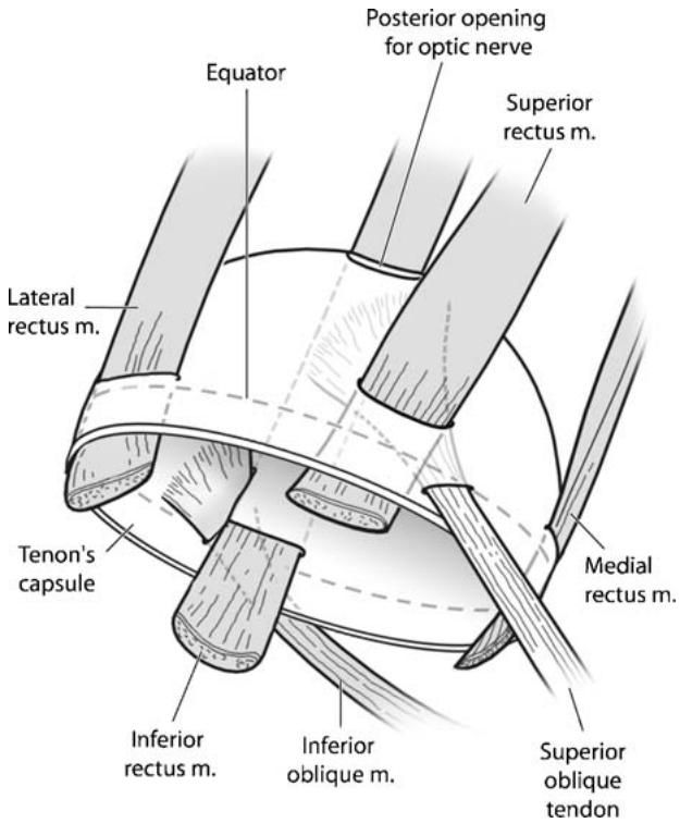
Fig. 1.9. Diagrammatic representation of Tenon’s capsule and relationship to the extraocular muscles. Note that the rectus muscles are located external to Tenon’s capsule posteriorly. They penetrate Tenon’s capsule to enter the episcleral space, then course anteriorly to insert on the sclera. The inferior oblique muscle and superior oblique tendon enter the episcleral space anteriorly and course posteriorly to insert on the sclera. The rectus muscle pulleys, muscle capsule, and intermuscular septum are not represented in this diagram
The potential space inside the superior oblique tendon sheath is continuous with the episcleral space. Numerous attachments extend from the sheath to adjacent structures including attachments to the global aspect of the sheath of the superior rectus muscle and attachments to the sheath of the levator palpebrae superioris muscle of the upper eyelid (>Fig. 1.10). Abnormalities involving this sheath often play a role in the etiology of Brown syndrome.
The fascial sheath of the inferior oblique muscle surrounds the muscle from its origin to insertion. It becomes thicker as the muscle approaches its insertion and it is usually tightly adherent to the orbital aspect of the sheath of the inferior rectus muscle. Small extensions of the sheath near the inferior oblique muscle insertion are directed to the sheath of the lateral rectus muscle and to the sheath surrounding the optic nerve posteriorly.