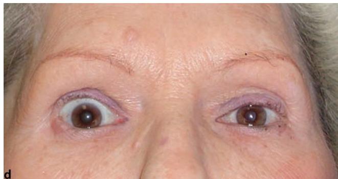
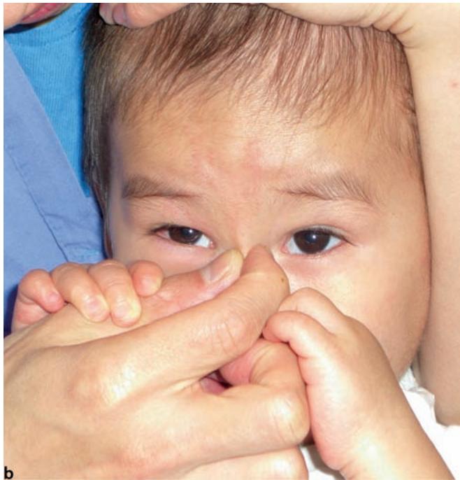
illusion of strabismus; c pseudoexotropia due to eyelid fissure asymmetry, and d pseudohypotropia due to right upper lid retraction.
1.1.5 Strabismus-Induced Eyelid Changes
One of the hallmarks of thyroid-related ophthalmopathy is retraction of one or more of the eyelids (>Fig. 1.5). While strabismus due to thyroid-related ophthalmopathy is generally obvious, subtle ocular motility disturbances can present. The presence of eyelid retraction may be the initial clue that the problem is due to thyroid-related ophthalmopathy, and can significantly alter key aspects of treatment including preoperative evaluation, timing of surgery, and the surgical plan itself.
1.3 The Sclera
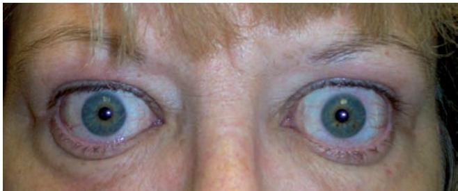
Fig. 1.5. Eyelid retraction in a patient with thyroid-related ophthalmopathy
1.1.6 Pseudoptosis
Patients with a large hypotropia, particularly those with a restrictive hypotropia, often present with a concurrent ptosis or pseudoptosis. An infant with a monocular elevator deficiency, for example, may appear to have concurrent, severe ptosis. When the child is made to fixate with the hypotropic eye, the apparent ptosis will resolve if the child can bring the hypotropic eye to the primary position, confirming the diagnosis of pseudoptosis in such cases. On the other hand, correction of the strabismus with surgery is often required to confirm a diagnosis of pseudoptosis if the child cannot bring the eye to the primary position (Chap. 27).
1.2 The Conjunctiva
The conjunctiva is a mucous membrane that covers the posterior surface of the eyelids and the anterior surface of the globe with the exception of the cornea. The bulbar conjunctiva merges with the stroma and epithelium of the cornea. Histologically, the conjunctiva is covered by nonkeratinized stratified squamous epithelial cells and has an underlying substantia propria. The bulbar conjunctiva overlies Tenon’s capsule. The conjunctiva contains numerous mucin-producing goblet cells, particularly in the fornix. The stroma consists of fragile connective tissue that contains lymphoid tissues. Accessory lacrimal glands are found in the conjunctiva of the upper and lower fornix. Though it is a single, continuous membrane, conceptually it is useful to divide the conjunctiva into two parts, namely the palpebral and bulbar conjunctivae. In general, the strabismus surgeon should never enter the palpebral conjunctiva, though an understanding of the anatomy and function of the palpebral conjunctiva as it relates to strabismus surgery remains important.
The palpebral conjunctiva begins on the lid margin at the mucocutaneous junction. The conjunctiva is tightly adherent to the underlying tarsus as it progresses into the fornix. The conjunctiva in the fornices is loose and is reflected into several folds, allowing movement of the globe not to be inhibited by connections between the palpebral and bulbar conjunctivae. The upper fornix is typically much deeper than the inferior fornix. A fat pad present in the inferior fornix should be identified and avoided during strabismus surgery (>Fig. 1.6; Chap. 25). Finally, from the fornices, the conjunctiva is reflected upon the globe where it is loosely adherent to the underlying Tenon’s fascia overlying the sclera, finally ending at the limbus.
There is some redundancy of the conjunctiva so that excision of small parts of the conjunctiva is well tolerated without altering its function and without significantly reducing its secretory capacity. Only the bulbar conjunctiva is routinely incised during strabismus surgery. Except for a small segment of conjunctiva immediately adjacent to the limbus, Tenon’s fascia will always be found deep to the bulbar conjunctiva. The lateral angle of the conjunctiva is rather nondescript and featureless compared to the medial angle of the conjunctiva. A fold of conjunctiva known as the plica semilunaris conjunctivae (referred to simply as plica here) is present in the medial angle of the conjunctiva and represents a fold in the bulbar conjunctiva. It serves no particular function in the human eye but corresponds to the nictitating membrane of some animal species. When malpositioned or accidentally incised during strabismus surgery, it can produce serious cosmetic and/or functional problems (Chap. 19). Another fold of conjunctiva near the medial canthus forms the caruncle, a transition zone between the conjunctiva and skin. It contains elements of skin and mucous membrane. Small lanugo hairs can often be seen growing out of its head. While the plica is essentially never intentionally incised or surgically altered during strabismus surgery, an incision through the caruncle may occasionally be used in the treatment of complex strabismus, such as the creation of a periosteal flap (Chap. 15) or repair of a lost muscle (Chap. 23).
1.3 The Sclera
The sclera is composed of densely packed collagen lamellae. It is continuous with the dura mater of the optic nerve. It also continues across the optic nerve head to form the lamina cribrosa. The sclera is penetrated by a variety of vascular and neural structures anteriorly and posteriorly. The thickness of the sclera also varies with age. It is typically thinnest in a newborn. The thickness of the sclera varies depending on its location on the globe. The sclera is thinnest behind the insertions of the rectus muscles, where its thickness is approximately 0.45 mm [9], a fact that is important to the strabismus surgeon since sutures may need to be passed into this thin sclera to recess a rectus muscle (>Fig. 1.7). The sclera is approximately 0.6–0.7 mm thick at the corneal limbus and 1.1–1.3 mm thick at the posterior pole [9] (>Fig. 1.8).
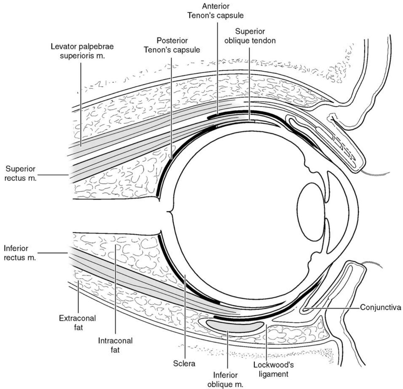
Fig. 1.6. Relationship of orbital fat to Tenon’s capsule and other structures. Violation of the posterior portions of Tenon’s capsule can result in intrusion of orbital fat into the surgical space