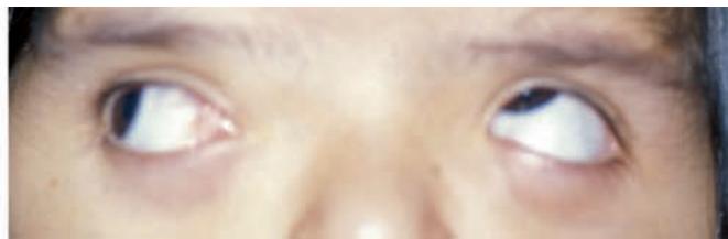
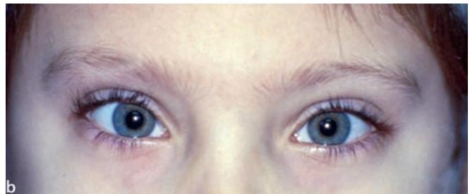
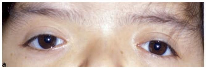
Fig. 1.1a,b. Lid fissure anatomy may provide initial clues in evaluating a patient with strabismus. a V-pattern strabismus and apparent inferior oblique overaction in a patient with down-slanting palpebral fissures. b A-pattern strabismus is common in patients with up-slanting palpebral fissures
1.1 The Ocular Adnexa
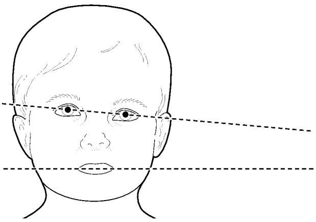
Fig. 1.2. Facial asymmetry in a patient with a history of early-onset superior oblique palsy. Note the midfacial hemihypoplasia on the side of the face toward the head tilt
Neurosurgical treatment of coronal synostosis may alter ocular alignment, changing the ultimate surgical plan, or even eliminating the need for strabismus surgery altogether.
1.1.4 Pseudostrabismus
Both normal and pathologic variations in eyelid fissure anatomy can produce the appearance of strabismus, despite normal alignment of the visual axis of the two eyes. In a pediatric ophthalmology practice, diagnosis of pseudoesotropia in infants with large epicanthal folds is probably the most common example (>Fig. 1.4a). Family members and primary care physicians alike may believe that strabismus is present because they do not see much “white” on the nasal aspect of the eye compared to the temporal aspect of the eye. Good advice to parents in this setting is to “ignore the white and look at the light,” pointing out the need to assess the corneal light reflex. After careful explanation, most parents can often recognize that the position and shape of the eyelids and other ocular adnexal structures can produce the appearance of strabismus, when in fact the eyes are aligned. It is sometimes difficult, however, to convince doubting parents that the eyes are straight. A simple demonstration of tightening the epicanthal folds by pinching the bridge of the nose can be an effective tool in convincing parents that the crossing that they see is an illusion (>Fig. 1.4b).
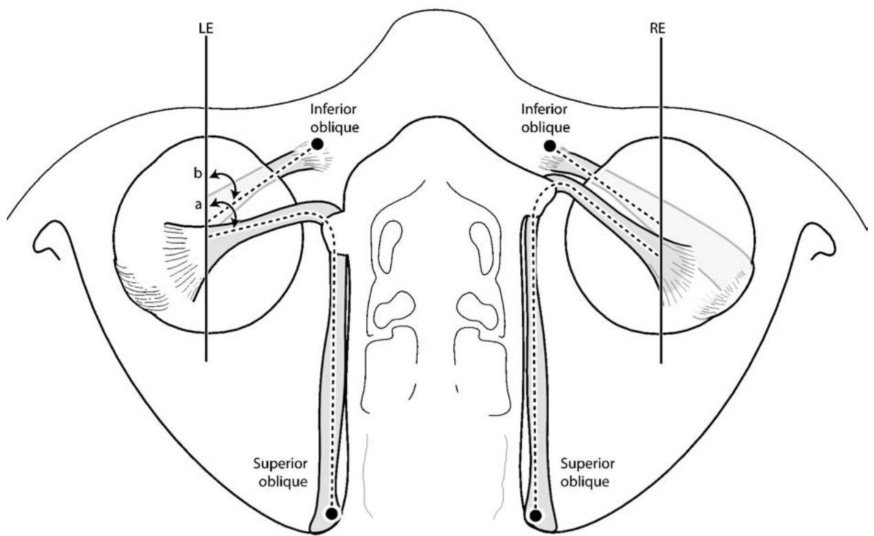
Fig. 1.3. Failure of the trochlea to advance anterior to the equator in a patient with unilateral coronal synostosis may result in reduction of depressing action on the globe with contraction of the superior oblique muscle

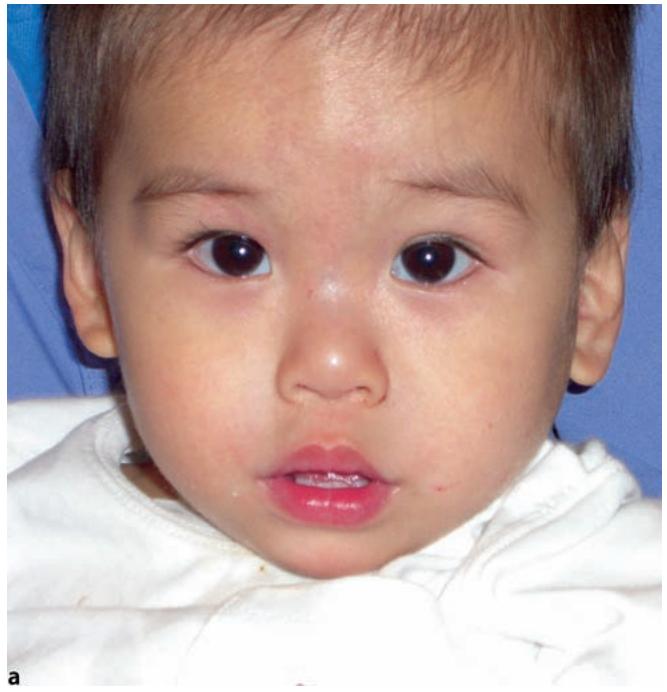
Fig. 1.4a–d. Pseudostrabismus: a pseudoesotropia, b with a simple demonstration to parents that the epicanthal folds are producing the
In the same way that prominent epicanthal folds can create the illusion of esotropia, abnormalities involving the lateral canthal area can create the illusion of exotropia. Temporal ptosis and dermatochalasis with prominent temporal hooding are just two examples of conditions that may produce the illusion of exotropia. An example of lid fissure asymmetry causing pseudoexotropia is shown in Fig. 1.4c. The illusion of vertical strabismus can be created by asymmetric ptosis and/or upper or lower eyelid retraction (>Fig. 1.4d). Similarly, changes in the lid fissures induced by strabismus surgery, especially when asymmetric, can be distressing to patients postoperatively (Chap. 26), both because of the presence of the eyelid asymmetry itself and because the patient may continue to believe that strabismus is still present, when in fact the eyes are actually well aligned.