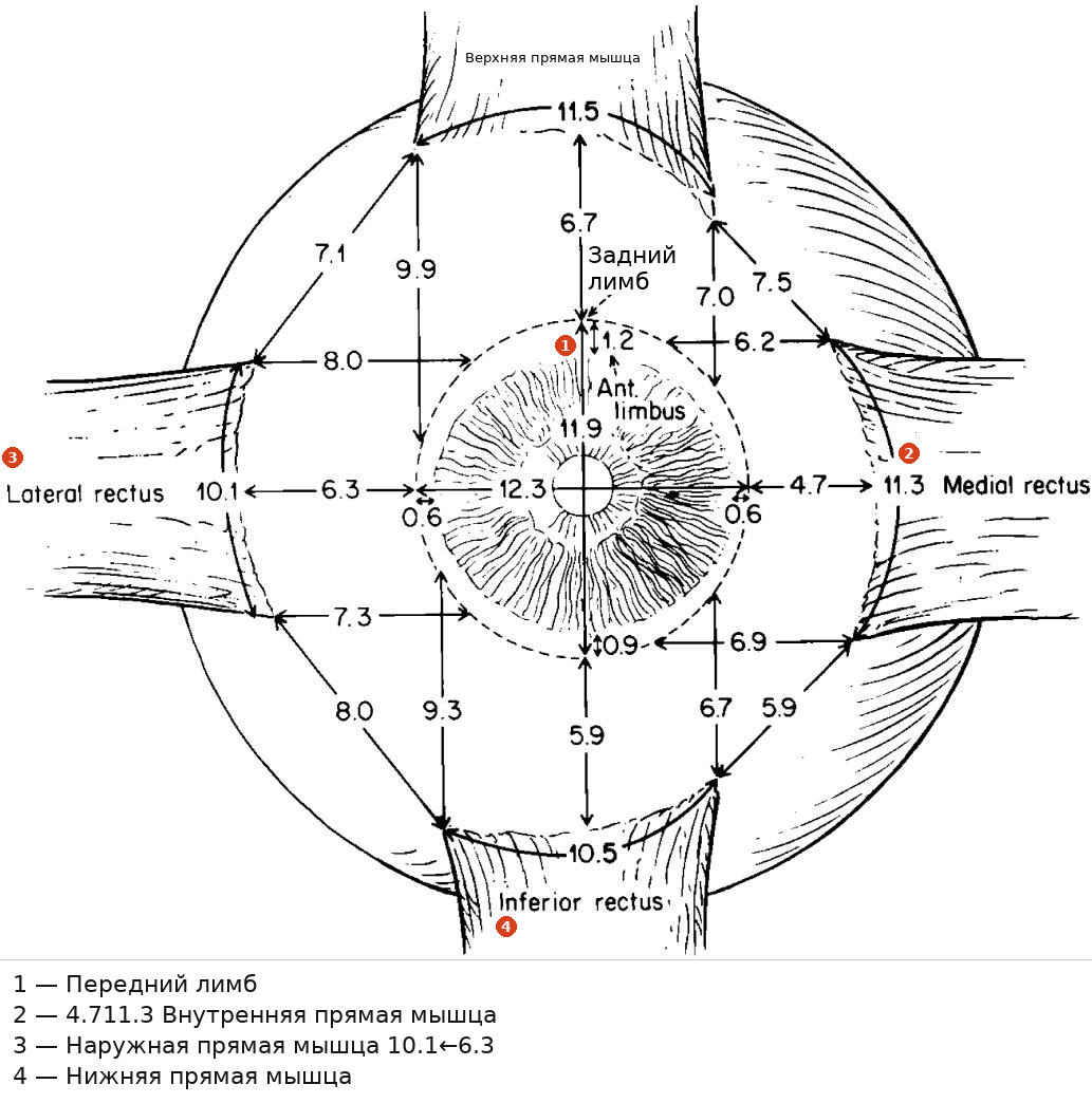
Рис. 1.12. Важные измерения размеров прямых мышц. Средняя ширина прикреплений прямых мышц, расстояние от прикрепления до лимба роговицы и длина сухожилия у взрослых. (С разрешения
Fig. 1.12. Important measurements of rectus muscle dimensions. Average width of rectus muscle insertions, distance of insertion to the corneal limbus and length of tendon, in adults. (With permission from
Apt L. An anatomical reevaluation of rectus muscle insertions. Trans Am Ophthalmol Soc 1980;78:365–375 [18])
Apt L. An anatomical reevaluation of rectus muscle insertions. Trans Am Ophthalmol Soc 1980;78:365–375 [18])
Таблица 1.1. Важные измерения размеров прямых мышц. Средняя ширина прикреплений прямых мышц у взрослого [18], расстояние от прикрепления до лимба роговицы [18] и длина сухожилия [26]
Table 1.1. Important measurements of rectus muscle dimensions. Average width of rectus muscle insertions in an adult [18], distance of insertion to the corneal limbus [18], and length of tendon [26]
| Мышца | Размеры сухожилия прямой мышцы | ||
| Расстояние от лимба (мм) | Ширина (мм) | Длина (мм) | |
| Внутренняя прямая мышца | 5.3 | 11.3 | 4 |
| Нижняя прямая мышца | 6.8 | 10.5 | 4.2 |
| Наружная прямая мышца | 6.9 | 10.1 | 6.65 |
| Верхняя прямая мышца | 7.9 | 11.5 | 5 |
| Muscle | Rectus muscle tendon dimensions | ||
| Distance from limbus (mm) | Width (mm) | Length (mm) | |
| Medial rectus | 5.3 | 11.3 | 4 |
| Inferior rectus | 6.8 | 10.5 | 4.2 |
| Lateral rectus | 6.9 | 10.1 | 6.65 |
| Superior rectus | 7.9 | 11.5 | 5 |
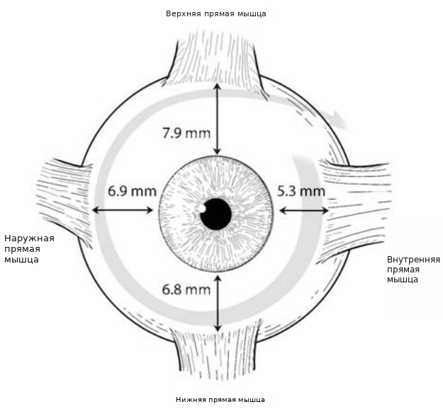
Рис. 1.13. Спираль Тильо
Fig. 1.13. The spiral of Tillaux

Рис. 1.14. Мышечные «подошвы» (слева от звездочки), представляющие собой мышечные пучки, перенаправленные позади места прикрепления, встречаются часто и, вероятно, не имеют функционального значения
Fig. 1.14. Muscle “footplates” (left of asterisk) representing muscle bundles redirected posterior to the insertion are common and are probably of no functional significance
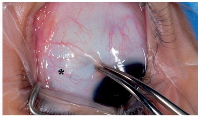
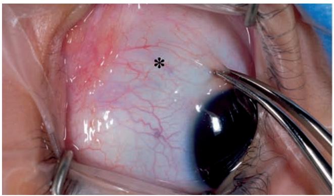
Рис. 1.15. Определение мест прикрепления прямых мышц путем визуализации их передних цилиарных сосудов под конъюнктивой при ротации глаза. Обратите внимание на движение передних цилиарных сосудов относительно конъюнктивальных сосудов
Fig. 1.15. Identifying the rectus muscle insertions by visualizing their anterior ciliary vessels beneath the conjunctiva as the eye is rotated. Note movement of the anterior ciliary vessels relative to the conjunctival vessels

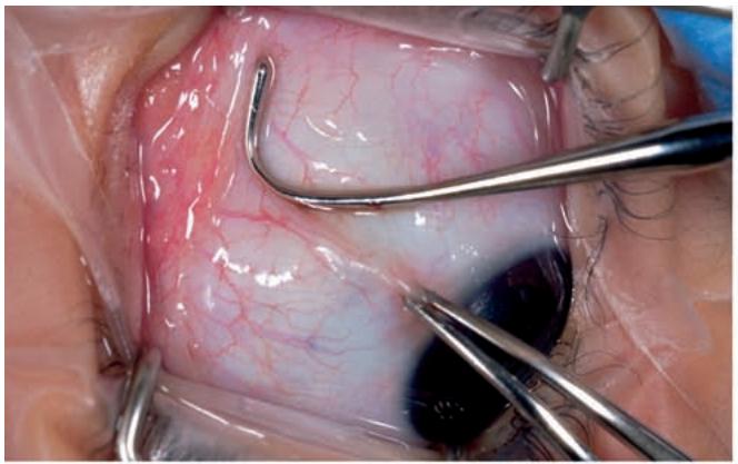
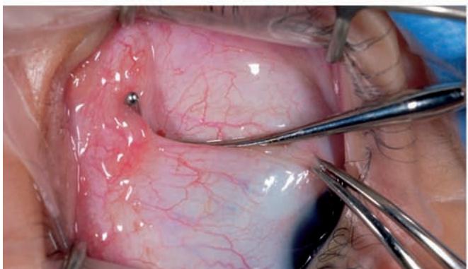
Рис. 1.16. Пальпация края прямой мышцы. После визуального определения места прикрепления крючок помещается рядом с краем мышцы (вверху слева). При надавливании крючком в сторону глазного яблока крючок перемещают в сторону мышцы (вверху справа). Видно, как мышца собирается у крючка по мере его продвижения (внизу)
Fig. 1.16. Palpation of a rectus muscle border. After visual identification of the insertion, a hook is placed adjacent to the muscle border (top left). While depressing the hook toward the globe, the hook is moved toward the muscle (top right). The muscle can be seen to bunch against the hook as it is advanced (bottom)
1.8 Кровоснабжение глазодвигательных мышц
1.8 Blood Supply to Extraocular Muscles
так называемые мышечные подошвы, которые когда-то считались имеющими значительное значение в этиологии косоглазия [19], сегодня представляются малозначимыми с функциональной точки зрения.
so-called muscle footplates, once thought to have considerable importance in the etiology of strabismus [19], appear today to be of little functional significance.
Прикрепления прямых мышц легко видны через неповрежденную конъюнктиву. Их расположение часто определяют в первую очередь по соответствующим передним цилиарным сосудам, которые выходят на эписклеру спереди от мест прикрепления прямых мышц. Эти сосуды легче всего идентифицировать при ротации глаза вперед-назад под прямым углом к ходу мышцы (>Рис. 1.15), где видно, что они перемещаются при ротации глазного яблока асинхронно по отношению к вышележащим конъюнктивальным сосудам. После идентификации сами мышцы обычно видны как слегка затемненные и немного приподнятые структуры под конъюнктивой.
The insertions of the rectus muscles can be easily seen through intact conjunctiva. Their locations are often first recognized by visualizing their associated anterior ciliary vessels as they course onto the episclera anterior to the rectus muscle insertions. These vessels are most readily identified as the eye is rotated to and fro at right angles to the path of the muscle (>Fig. 1.15) where they can be seen to move with globe rotation asynchronously from the overlying conjunctival vessels. Once identified, the muscles themselves can usually be seen as slightly darkened and slightly raised structures beneath the conjunctiva.