1.5 Система блоков прямых мышц
1.5 The Rectus Muscle Pulley System
Отражение, исходящее от капсулы Тенона, охватывает заднюю часть глазодвигательных мышц, которые в данной области орбиты находятся вне капсулы. Фиброэластические футляры, состоящие из плотных пучков коллагена, эластина и гладких мышц, окружают прямые мышцы. Эти футляры подвешены к орбите и футлярам соседних глазодвигательных мышц с помощью тканевых тяжей аналогичного состава. Эти футляры и их связи со стенками орбиты, которые часто называют удерживающими связками, выполняют значительно более сложную функцию, чем просто «удержание» движения глазного яблока, и термин «удерживающие связки», вероятно, следует исключить. Конденсации и расширения этих мышечных оболочек в конечном итоге связаны с соединениями и в переднем отделе. Некоторые рассматривают все эти структуры совместно как капсулу Тенона [11], хотя для практических целей, несмотря на то что они могут быть непрерывными и постепенно переходить одна в другую, блоки прямых мышц настолько специализированы по функциям, что их следует рассматривать отдельно от капсулы Тенона.
A reflection extending from Tenon’s capsule envelops the posterior portion of the extraocular muscles that are extrinsic to the capsule at this position in the orbit. Fibroelastic sleeves consisting of dense bands of collagen, elastin, and smooth muscle surround the rectus muscles. These sleeves are suspended from the orbit and adjacent extraocular muscle sleeves by bands of tissue having similar composition. Often referred to as check ligaments, these sleeves and their connections to the orbital walls have a significantly more complex function than simply to “check” movement of the globe and the term check ligaments should probably be discarded. Condensations and extensions from these muscle sheaths ultimately are associated with connections anteriorly as well. Some consider all of these structures collectively as Tenon’s capsule [11] though for practical purposes while they may all be continuous, with one blending gradually into the other, the rectus muscle pulleys are so specialized in function that they should be considered separate from Tenon’s capsule.
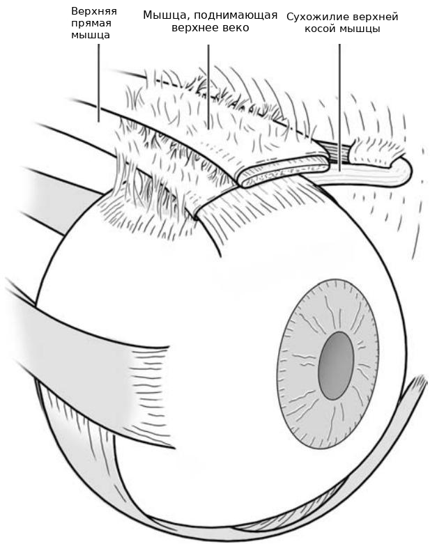
Рис. 1.10. Сложная оболочка сухожилия верхней косой мышцы. Обратите внимание на слабые прикрепления к оболочке верхней прямой мышцы и к мышце, поднимающей верхнее веко
Fig. 1.10. Complex sheath of the superior oblique tendon. Note tenuous attachments to the sheath of the superior rectus muscle and to the levator muscle of the upper eyelid
Компьютерная томография высокого разрешения и магнитно-резонансная томография продемонстрировали, что ход прямых мышц остается стабильным относительно прилегающих стенок орбиты на протяжении большей части их пути в орбите, даже при движениях глаз и после обширных хирургических процедур по транспозиции [12,13]. Только передний отдел мышц фактически перемещается при нормальных движениях глаз в положениях вторичного взора, в то время как задние отделы прямых мышц относительно фиксированы в положении блоками прямых мышц, которые частично расположены вблизи экватора глазного яблока.
High-resolution computed tomography and magnetic resonance imaging have demonstrated that the paths of the rectus muscles remain stable relative to their adjacent orbital walls throughout most of their course in the orbit, even during eye movements and following large surgical transposition procedures [12,13]. Only the anterior aspect of the muscles actually moves during normal eye movements into secondary gaze positions, while the posterior aspects of the rectus muscles are relatively fixed in position by rectus muscle pulleys which are in part located near the equator of the globe.
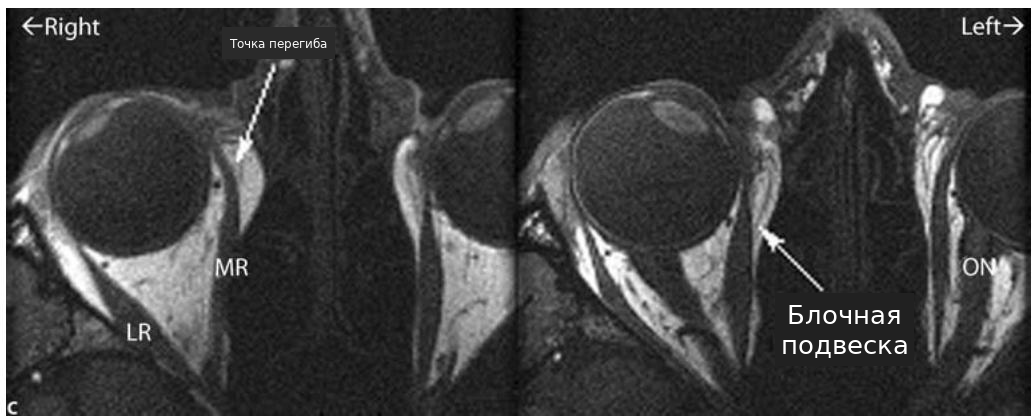
Блоки прямых мышц по сути функционируют как эффективные места начала прямых мышц [14] (>Рис. 1.11). Эти мышечные футляры непрерывно переходят в капсулу Тенона спереди и сзади. Блоки имеют важное значение для изменения как хода прямых мышц через орбиту, так и их функции. Эти футляры расположены вблизи экватора глазного яблока и имеют размер в передне-заднем направлении примерно 13–19 мм [13]. При сокращении мышцы футляры действуют как блоки, ограничивая ход прямых мышц и перенаправляя силу сокращения к глазному яблоку. Было показано, что орбитальный слой прямых глазодвигательных мышц прикрепляется к соответствующему блоку прямой мышцы, а не к глазному яблоку [15]. Гипотеза активного блока, предложенная Demer и соавторами [15], предполагает, что глобальный слой каждой прямой глазодвигательной мышцы вращает глазное яблоко, в то время как орбитальный слой мышцы прикрепляется к соответствующему блоку и влияет на ось вращения прямой мышцы за счет изменения положения блока при движениях глазного яблока (>Рис. 1.11c). Считается, что такое устройство дает механическое объяснение важных аспектов движений глаз, включая закон Листинга [15,16].
The rectus muscle pulleys essentially function as the effective origins of the rectus muscles [14] (>Fig. 1.11). These muscle sleeves are continuous with Tenon’s capsule anteriorly and posteriorly. The pulleys are important for altering both the paths of the rectus muscles through the orbit and their function. These sleeves are located near the equator of the globe and are approximately 13–19 mm in anterior–posterior dimension [13]. During muscle contraction, the sleeves act as pulleys, restraining the rectus muscles paths and re-directing the force of contraction toward the globe. The orbital layer of the rectus extraocular muscles has been demonstrated to insert into the corresponding rectus muscle pulley, rather than on the globe [15]. The active-pulley hypothesis proposed by Demer and coworkers [15] suggests that the global layer of each rectus extraocular muscle rotates the globe while the orbital layer of the muscle inserts on its respective pulley and influences the rotational axis of the rectus muscle through changes in the position of the pulley during movements of the globe (>Fig. 1.11c). This arrangement is believed to provide a mechanical explanation of important aspects of eye movements including Listing’s law [15,16].
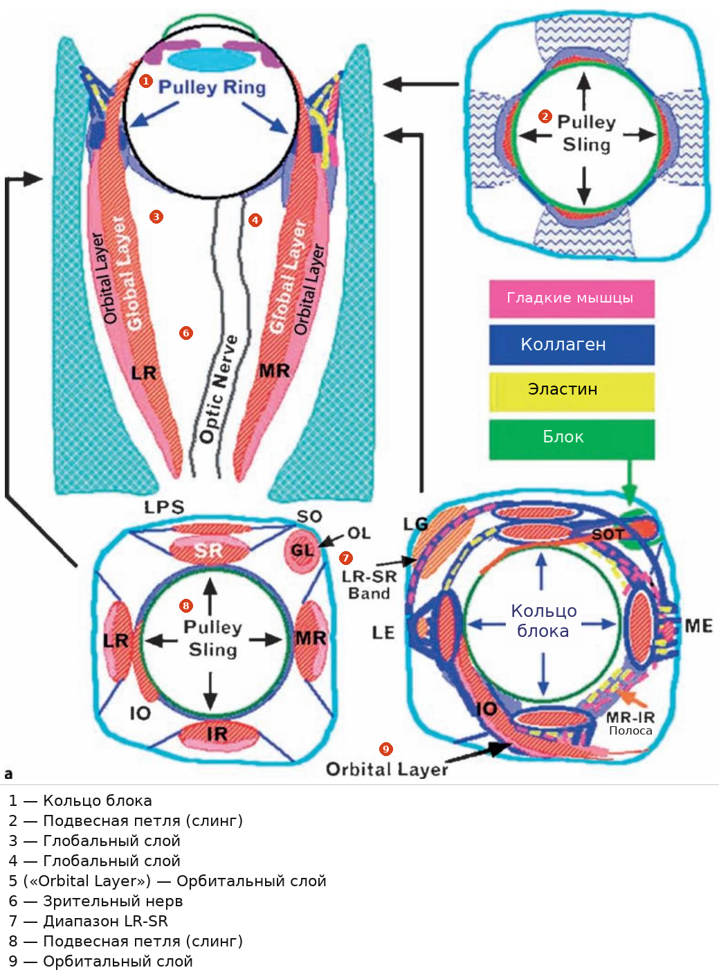
Рис. 1.11a–c. Блоки прямых мышц. a Схема блоков прямых мышц. (GL глобальный слой, IO нижняя косая мышца, IR нижняя прямая мышца, LE латеральный энтезис, LG слезная железа, LPS мышца, поднимающая верхнее веко, LR лат-
Fig. 1.11a–c.Rectus muscle pulleys. a Schematic of rectus muscle pulleys. (GL Global layer, IO inferior oblique, IR inferior rectus, LE lateral enthesis, LG lacrimal gland, LPS levator palpebrae superioris, LR lat-
еральная прямая мышца, ME медиальный энтезис, MR внутренняя прямая мышца, OL орбитальный слой, SO верхняя косая мышца, SR верхняя прямая мышца)
eral rectus, ME medial enthesis, MR medial rectus, OL orbital layer, SO superior oblique, SR superior rectus)
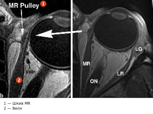
Рис. 1.11a–c. (продолжение) Блоки прямых мышц. b Магнитно-резонансная томография, демонстрирующая положение блока внутренней прямой мышцы. c Изменение положения блока, происходящее при сокращении мышцы. {a С разрешения Demer JL (2006) Current concepts of mechanical and neural factors in ocular motility. Curr Opin Neurol 19: 4–13; copyright 2006 Lippincott Williams and Wilkins [16]; b и c предоставлено Joseph L Demer, MD; грант NIH EY08313}W
Fig. 1.11a–c. (continued) Rectus muscle pulleys. b Magnetic resonance imaging demonstrating position of pulley for medial rectus muscle. c Change in pulley position that occurs during muscle contraction. {a With permission from Demer JL (2006) Current concepts of mechanical and neural factors in ocular motility. Curr Opin Neurol 19: 4–13; copyright 2006 Lippincott Williams and Wilkins [16]; b and c courtesy of Joseph L Demer, MD; NIH grant EY08313}W
1.6 Макроанатомия глазодвигательных мышц
1.6 Gross Anatomy of the Extraocular Muscles
1.6.1 Прямые мышцы
1.6.1 Rectus Muscles
Каждая из четырех прямых мышц берет начало в заднем отделе орбиты у кольца Цинна, окружающего зрительный канал и нижнюю часть верхней глазничной щели. Считается, что фасциальные сращения между местами прикрепления внутренней и верхней прямых мышц с твердой оболочкой, покрывающей зрительный нерв, являются причиной боли при движениях глаз у пациентов с острым зрительным нейритом. Место прикрепления наружной прямой мышцы имеет верхнюю и нижнюю головки, расположенные по разные стороны от верхней глазничной щели [17]. Для практических целей можно считать, что длина прямых мышц у взрослого составляет примерно 40 мм. Проходя вперед от кольца Цинна, внутренняя, наружная и нижняя прямые мышцы на значительной части своей длины следуют вдоль прилегающих стенок орбиты, в то время как верхняя прямая мышца отделена от свода орбиты мышцей, поднимающей верхнее веко.
Each of the four rectus muscles originates in the posterior orbit at the annulus of the Zinn surrounding the optic canal and the inferior portion of the superior orbital fissure. Fascial attachments between the origins of the medial and superior rectus muscles into the dura covering the optic nerve are thought to be the cause of pain with eye movements in patients with acute optic neuritis. The origin of the lateral rectus muscle has a superior and inferior head that are located on opposite sides of the superior orbital fissure [17]. For practical purposes, the rectus muscles can be considered to be approximately 40 mm in length in an adult. Coursing anteriorly from the annulus of Zinn, the medial, lateral, and inferior rectus muscles follow the course of adjacent orbital walls for a good portion of their length, while the superior rectus muscle is separated from the orbital roof by the levator palpebrae superioris muscle of the upper eyelid.
Пути прямых мышц в орбите резко изгибаются в сторону глазного яблока, начиная примерно за 7–10 мм до экватора, так как вышеописанные соединительнотканные/мышечные блоки изменяют их направление. Тонкие прямые мышцы в конечном итоге проникают сквозь капсулу Тенона за 7–10 мм до их прикрепления к склере. После вхождения в эписклеральное пространство и дальнейшего продвижения вперед каждая из них становится сухожильной и в итоге прикрепляется к склере в виде сухожилия позади лимба. Сухожильные прикрепления каждой из прямых мышц имеют ширину примерно 10–11 мм [18] (>Таблица 1.1).
The paths of the rectus muscles in the orbit curve sharply toward the globe starting approximately 7–10 mm from the equator as the connective tissue/muscular pulleys described above alter their paths. The thin rectus muscles eventually penetrate Tenon’s capsule 7–10 mm from their insertions into the sclera. After entering the episcleral space and coursing further anteriorly, each eventually becomes tendinous, ultimately inserting into the sclera as a tendon posterior to the limbus. The tendinous insertions of each of the rectus muscles are roughly 10–11 mm in width [18] (>Table 1.1).
Среднее расстояние от прикрепления каждой прямой мышцы до лимба показано в Таблице 1.1 и на Рис. 1.12 вместе с другими ключевыми анатомическими особенностями. Прикрепление внутренней прямой мышцы обычно расположено ближе всего к лимбу, далее следуют нижняя прямая, наружная прямая и, наконец, верхняя прямая мышца, которая обычно прикрепляется дальше всего от лимба. Места прикрепления, особенно вертикальных прямых мышц, изогнуты выпуклостью от лимба. Височные края вертикальных прямых мышц расположены дальше от лимба, чем носовые края. Отклонения от указанных средних значений встречаются часто. Окружность, соединяющая центры прикреплений прямых мышц, известна как спираль Тильо (>Рис. 1.13). Спираль Тильо имеет несколько важных значений: (1) помогает хирургу сохранять ориентацию во время операции и гарантировать, что операция проводится на нужной мышце, (2) дает представление об объеме и типе ранее проведенных операций по поводу косоглазия при повторных вмешательствах и (3) служит ориентиром для правильного повторного прикрепления транспонированных и смещенных вперед мышц. Нередко небольшие пучки мышечных волокон перенаправляются назад вблизи места прикрепления и фиксируются на несколько миллиметров позади остальных прикреплений прямых мышц (>Рис. 1.14). Эти 1.6 Макроанатомия глазодвигательных мышц
The average distance of each rectus muscle insertion from the limbus is shown in Table 1.1 and Fig. 1.12, along with other key anatomical features. The medial rectus muscle insertion is typically located closest to the limbus, followed by the inferior rectus, lateral rectus, and finally the superior rectus muscle, which is typically inserted furthest from the limbus. The insertions, particularly those of the vertical rectus muscles, are curved with their convexity away from the limbus. The temporal corners of the vertical rectus muscles are further from the limbus than are the nasal corners. Variations from the means shown are common. A circular line connecting the center of the rectus muscle insertions is known as the spiral of Tillaux (>Fig. 1.13). The spiral of Tillaux has several important implications: (1) helping the surgeon to remain oriented during surgery, and helping to assure surgery is performed on the correct muscle, (2) providing some insight as to the amount and type of previous strabismus surgery performed during reoperations, and (3) providing landmarks to help guide reinsertion of transposed and advanced muscles. It is not uncommon for small bundles of muscle fibers to be re-directed posteriorly near the insertion to insert several millimeters behind the remainder of the rectus muscle insertions (>Fig. 1.14). These 1.6 Gross Anatomy of the Extraocular Muscles