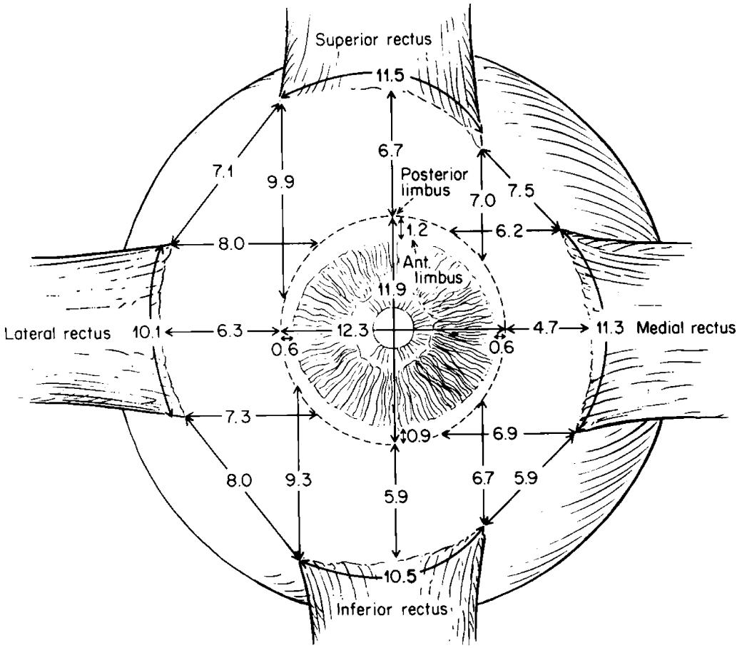
Fig. 1.12. Important measurements of rectus muscle dimensions. Average width of rectus muscle insertions, distance of insertion to the corneal limbus and length of tendon, in adults. (With permission from
Apt L. An anatomical reevaluation of rectus muscle insertions. Trans Am Ophthalmol Soc 1980;78:365–375 [18])
Table 1.1. Important measurements of rectus muscle dimensions. Average width of rectus muscle insertions in an adult [18], distance of insertion to the corneal limbus [18], and length of tendon [26]
| Muscle | Rectus muscle tendon dimensions | ||
| Distance from limbus (mm) | Width (mm) | Length (mm) | |
| Medial rectus | 5.3 | 11.3 | 4 |
| Inferior rectus | 6.8 | 10.5 | 4.2 |
| Lateral rectus | 6.9 | 10.1 | 6.65 |
| Superior rectus | 7.9 | 11.5 | 5 |
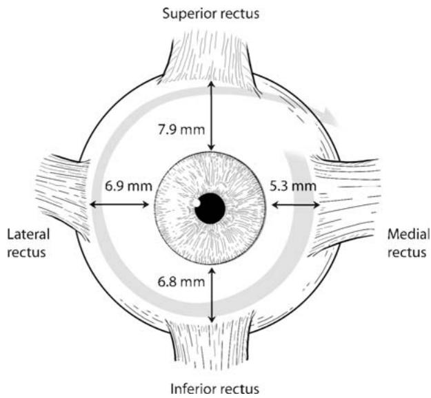
Fig. 1.13. The spiral of Tillaux

Fig. 1.14. Muscle “footplates” (left of asterisk) representing muscle bundles redirected posterior to the insertion are common and are probably of no functional significance
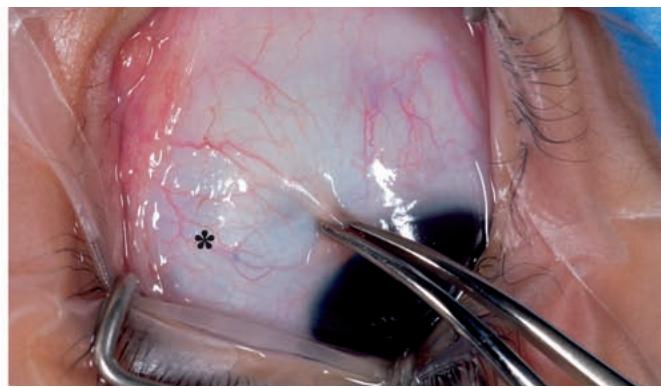
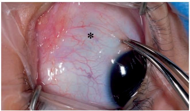
Fig. 1.15. Identifying the rectus muscle insertions by visualizing their anterior ciliary vessels beneath the conjunctiva as the eye is rotated. Note movement of the anterior ciliary vessels relative to the conjunctival vessels

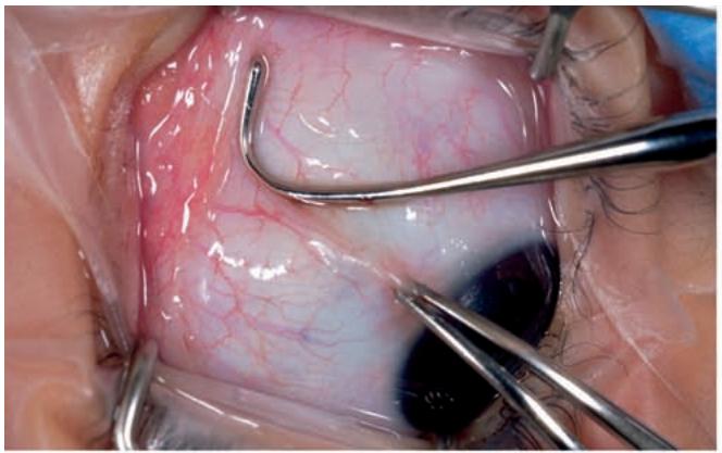
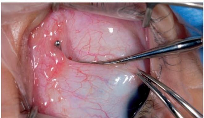
Fig. 1.16. Palpation of a rectus muscle border. After visual identification of the insertion, a hook is placed adjacent to the muscle border (top left). While depressing the hook toward the globe, the hook is moved toward the muscle (top right). The muscle can be seen to bunch against the hook as it is advanced (bottom)
1.8 Blood Supply to Extraocular Muscles
so-called muscle footplates, once thought to have considerable importance in the etiology of strabismus [19], appear today to be of little functional significance.
The insertions of the rectus muscles can be easily seen through intact conjunctiva. Their locations are often first recognized by visualizing their associated anterior ciliary vessels as they course onto the episclera anterior to the rectus muscle insertions. These vessels are most readily identified as the eye is rotated to and fro at right angles to the path of the muscle (>Fig. 1.15) where they can be seen to move with globe rotation asynchronously from the overlying conjunctival vessels. Once identified, the muscles themselves can usually be seen as slightly darkened and slightly raised structures beneath the conjunctiva.