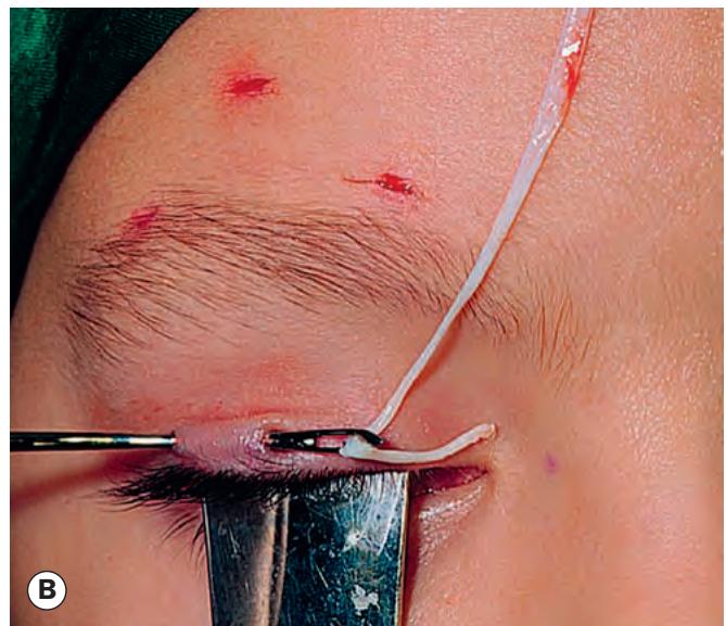
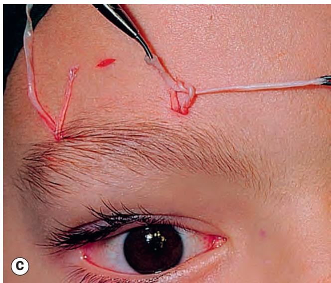
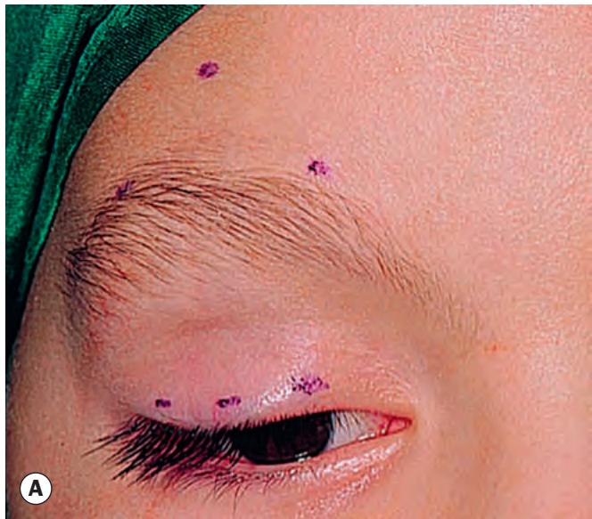
Fig. 1.60 Brow suspension. (A) Site of incisions marked; (B) threading of fascia lata strips; (C) tightening and tying of strips
(Courtesy of AG Tyers and JRO Collin, from Colour Atlas of Ophthalmic Plastic Surgery, Butterworth-Heinemann 2001)
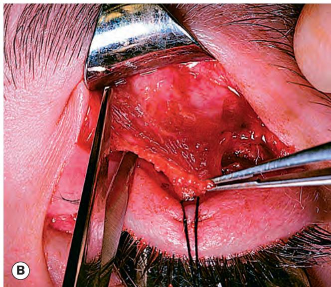
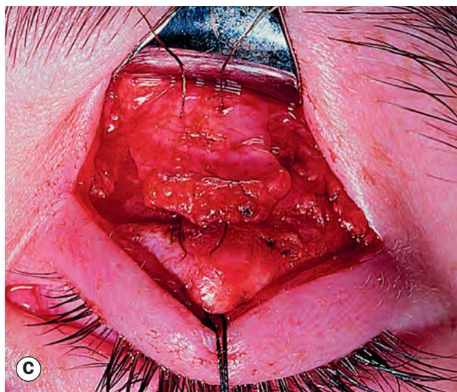
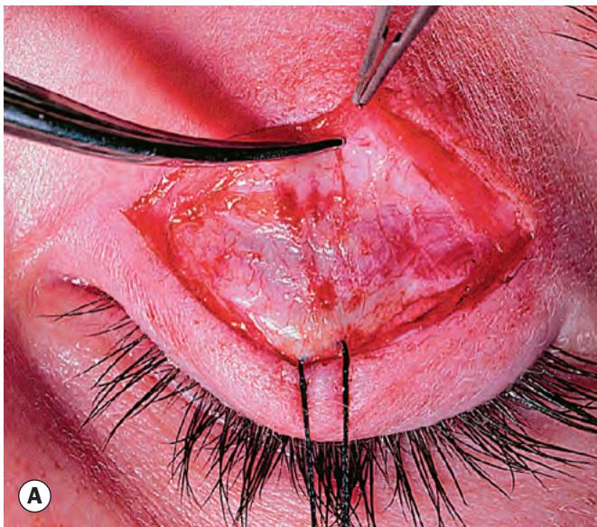
Fig. 1.59 Anterior levator resection. (A) Skin incision;
(B) dissection and resection of levator aponeurosis;
(C) levator reattachment to the tarsal plate
(Courtesy of AG Tyers and JRO Collin, from Colour Atlas of Ophthalmic Plastic Surgery, Butterworth-Heinemann 2001)
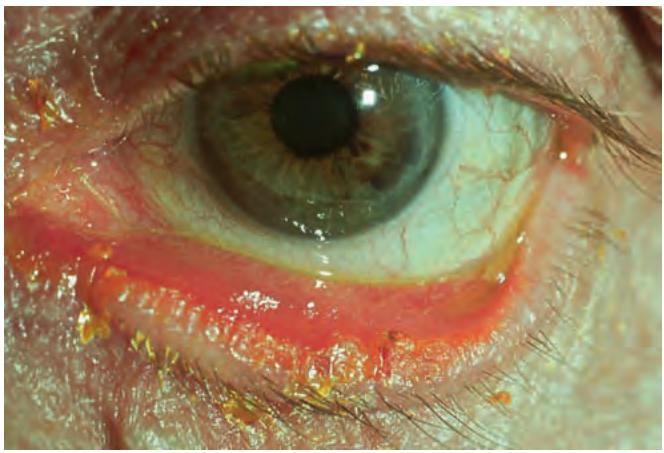
Fig. 1.61 Severe long-standing involutional ectropion with keratinization of the marginal conjunctiva
(Courtesy of C Barry)
Lateral canthal tendon laxity, characterized by a rounded appearance of the lateral canthus (see Fig. 1.46B) and the ability to pull the lower lid medially more than 2 mm.
Medial canthal tendon laxity, demonstrated by pulling the lower lid laterally and observing the position of the inferior punctum. If the lid is normal the punctum should not be displaced more than 1–2 mm. If laxity is mild the punctum reaches the limbus, and if severe it may reach the pupil.
Treatment
The approach to repair depends on apparent causation and the predominant location of the ectropion.
Generalized ectropion is treated with repair of horizontal lid laxity. This is achieved with a lateral tarsal strip procedure, in which the lower canthal tendon is tightened by shortening and reattachment to the lateral orbital rim (Fig. 1.62); this is particularly helpful if the lateral canthus is rounded and lax, with associated tear overflow. Excision of a tarsoconjunctival pentagon (Fig. 1.63) is an alternative that can be placed to excise an area of misdirected lashes or keratinized conjunctiva.
Medial ectropion, if mild, may be treated with a medial conjunctival diamond excision (medial spindle procedure), though must often be combined with a tarsal strip or lateral canthal sling (see “Treatment”), or pentagon excision as significant horizontal laxity frequently co-exists.
Medial canthal tendon laxity, if marked, requires stabilization prior to horizontal shortening to avoid excessive dragging of the punctum laterally.
Punctal ectropion without more extensive lid involvement is considered in Ch. 2.