

Fig. 1.35 Cryotherapy to the eyelid in distichiasis. (A) Separation of the anterior and posterior lamellae; (B) application of cryoprobe to the posterior lamella
(Courtesy of AG Tyers and JRO Collin, from Colour Atlas of Ophthalmic Plastic Surgery, Butterworth-Heinemann 2001)
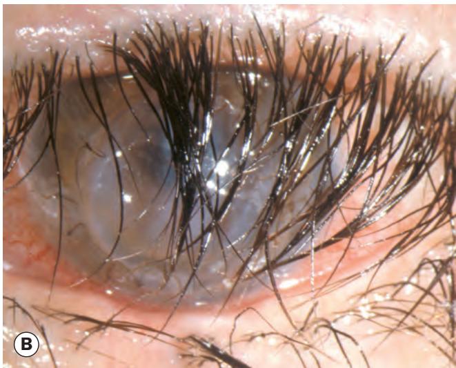


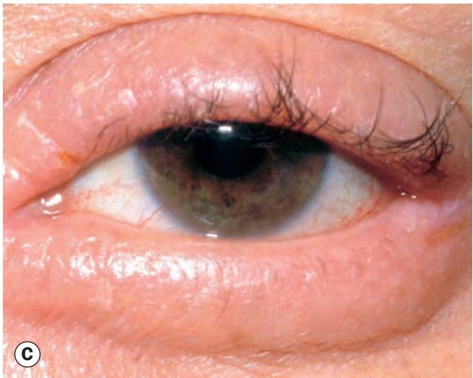
Fig. 1.36 Miscellaneous eyelash disorders. (A) Eyelash ptosis; (B) trichomegaly; (C) madarosis; (D) poliosis
(Courtesy of A Pearson – fig. A; L Merin – fig. B; S Tuft – fig. C)
Allergic Disorders
Table 1.2 Cause of madarosis
| 1. Local Chronic anterior lid margin disease Infiltrating lid tumours Burns Radiotherapy or cryotherapy of lid tumours |
| 2. Skin disorders Generalized alopecia Psoriasis |
| 3. Systemic diseases Myxoedema Systemic lupus erythematosus Acquired syphilis Lepromatous leprosy |
| 4. Following removal Procedures for trichiasis Trichotillomania – psychiatric disorder of hair removal |
Table 1.3 Causes of poliosis
| 1. Ocular Chronic anterior blepharitis Sympathetic ophthalmitis Idiopathic uveitis |
| 2. Systemic Vogt–Koyanagi–Harada syndrome Waardenburg syndrome Vitiligo Marfan syndrome Tuberous sclerosis |
ALLERGIC DISORDERS
Acute allergic oedema
Acute allergic oedema is usually caused by exposure to pollen or by insect bites, and manifests with the sudden onset of bilateral boggy periocular oedema (Fig. 1.37A), often accompanied by conjunctival swelling (chemosis – see Ch. 5). Treatment is often unnecessary, but systemic antihistamines are sometimes given.
Contact dermatitis
Contact dermatitis is an inflammatory response that usually follows exposure to a medication such as eye drops (often preservative-containing), cosmetics or metals. An irritant can also cause a non-allergic toxic dermatitis. The individual is sensitized on first exposure and develops an immune reaction on further exposure; the mediating reaction is type IV (delayed type) hypersensitivity. Signs consist of lid skin scaling, angular fissuring, oedema and tightness (Fig. 1.37B); there may be chemosis, redness and papillary conjunctivitis. Corneal involvement is usually limited to punctate epithelial erosions. Treatment consists primarily of avoidance of allergen exposure, provided it can be identified. Cold compresses provide symptomatic relief. Topical steroids and oral antihistamines can be used, but are rarely required.