Surgical excision
Surgical excision aims to remove the entire tumour with preservation of as much normal tissue as possible. Smaller tumours can be removed via an excision biopsy and the defect closed directly, whilst awaiting histological confirmation of complete clearance. Most small BCCs can be cured by excision of the tumour together with a 2–4 mm margin of clinically normal tissue. More radical surgical excision is required for large BCCs and aggressive tumours such as SCC, SGC and melanoma. It may not be possible to close all defects at the time of initial removal, but it is necessary to ensure complete clearance of tumour prior to undertaking any reconstruction. There are several options for the coordination of histopathological diagnosis and tumour clearance with excision.
Conventional paraffin-embedded specimen. Rapid processing can reduce the interval to confirmation of histological clearance but still requires that reconstruction be performed as a separate procedure. Faster confirmation can be achieved using either frozen-section control or micrographic surgery (see next), and reconstruction can then take place on the same day.
Standard frozen section involves histological examination of the margins of the excised specimen at the time of surgery to ensure they are tumour-free. If no tumour cells are detected, the eyelid is reconstructed on the same day; if residual tumour is present, further excision is performed at the appropriate edge of the surgical site until no tumour is detected.
Mohs micrographic surgery involves layered excision of the tumour; specimens are usually examined frozen. Processing of each layer enables a map of the edges of the tumour to be

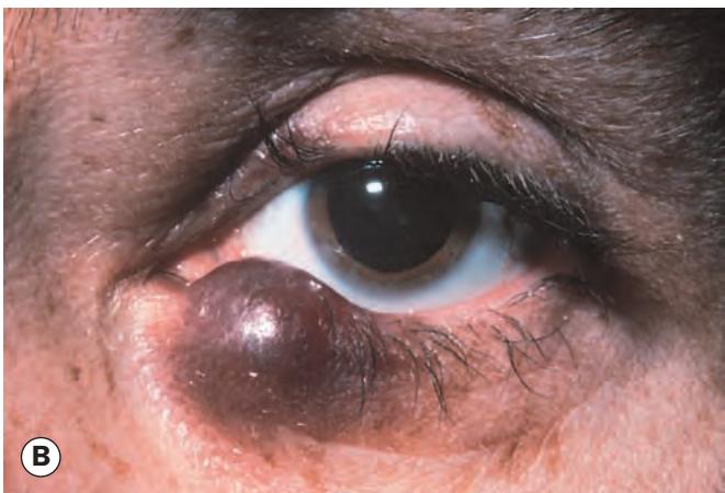
Fig. 1.27 Kaposi sarcoma. (A) Histopathology shows a proliferation of predominantly spindle-shaped cells; vascular channels are evident; (B) clinical appearance
(Courtesy of J Harry – fig. A)
developed. Further tissue is taken in any area where tumour is still present until clearance is achieved. Although time-consuming, this technique maximizes the chances of total tumour excision whilst minimizing sacrifice of normal tissue. This is a particularly useful technique for tumours that grow diffusely and have indefinite margins with finger-like extensions, such as sclerosing BCC, SCC, recurrent tumours and those involving the medial or lateral canthi. The irregular contours around the eyelids and extension of tumours into orbital fat can make interpretation difficult.
Reconstruction
The technique of reconstruction depends on the extent of tissue removed. It is important to reconstruct both anterior and posterior lamellae, each of which must be reconstructed with similar tissue. Anterior lamellar defects may be closed directly or with a local flap or skin graft. Options for the repair of full-thickness defects are set out below.
Small defects involving less than one-third of the eyelid can usually be closed directly, provided the surrounding tissue is sufficiently elastic to allow approximation of the cut edges (Fig. 1.28). If necessary, a lateral cantholysis can be performed for increased mobilization.
Moderate size defects involving up to half of the eyelid may require a flap (e.g. Tenzel semicircular) for closure (Fig. 1.29).
Large defects involving over half of the eyelid may be closed by one of the following techniques:
☐ Posterior lamellar reconstruction may involve an upper lid free tarsal graft, buccal mucous membrane or hard palate graft, or a Hughes tarsoconjunctival flap from the upper lid, which is left attached for 4–6 weeks before transection (Fig. 1.30).
☐ Anterior lamellar reconstruction may involve skin advancement, a local skin flap or a free skin graft (Fig. 1.31); the patient must be made aware that grafted skin is unlikely to be a perfect match. At least one reconstructed lamella requires its own blood supply to maximize the viability of a free graft component.
Laissez-faire
Full reconstruction of the defect created by tumour removal may not always be required. In the laissez-faire approach the wound edges are approximated as far as possible and the defect is allowed to granulate and heal by secondary intention. Even large defects can often achieve a satisfactory outcome with time.
Radiotherapy
The recurrence rate following irradiation alone is higher than after surgery, and radiotherapy does not allow histological confirmation of tumour eradication. Recurrences following radiotherapy are difficult to treat surgically because of the poor healing properties of irradiated tissue. However, it still has utility in some circumstances.
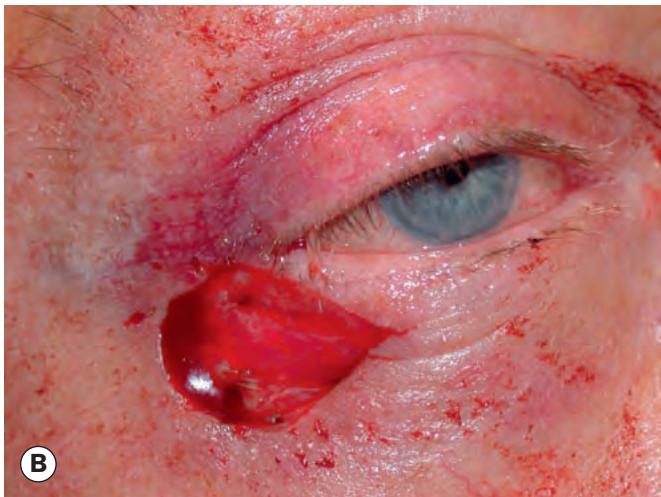
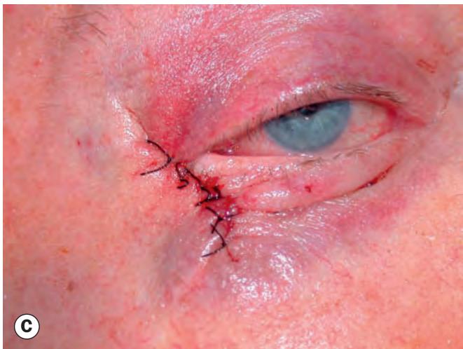
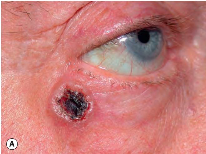
Fig. 1.28 Direct closure. (A) Preoperative appearance of a basal cell carcinoma; (B) appearance following excision; (C) direct closure of defect
(Courtesy of A Pearson)