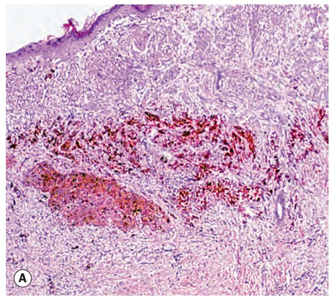
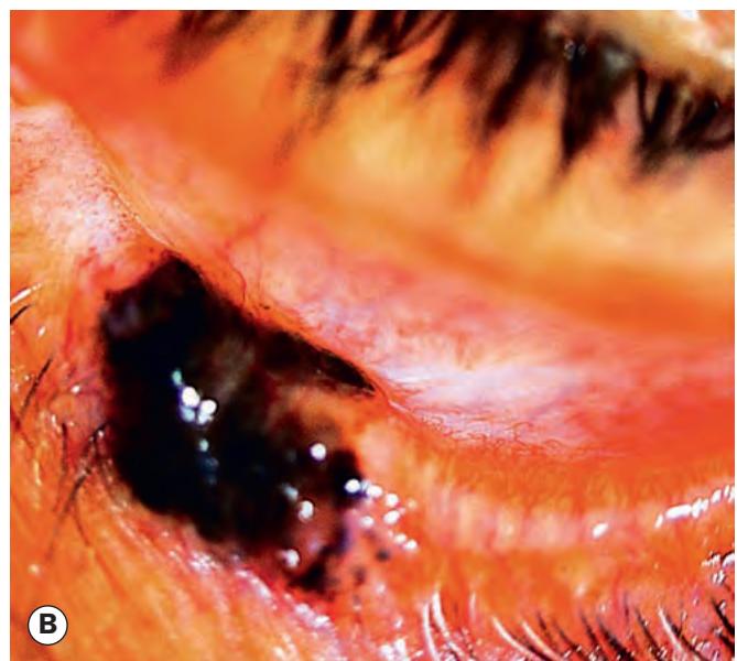
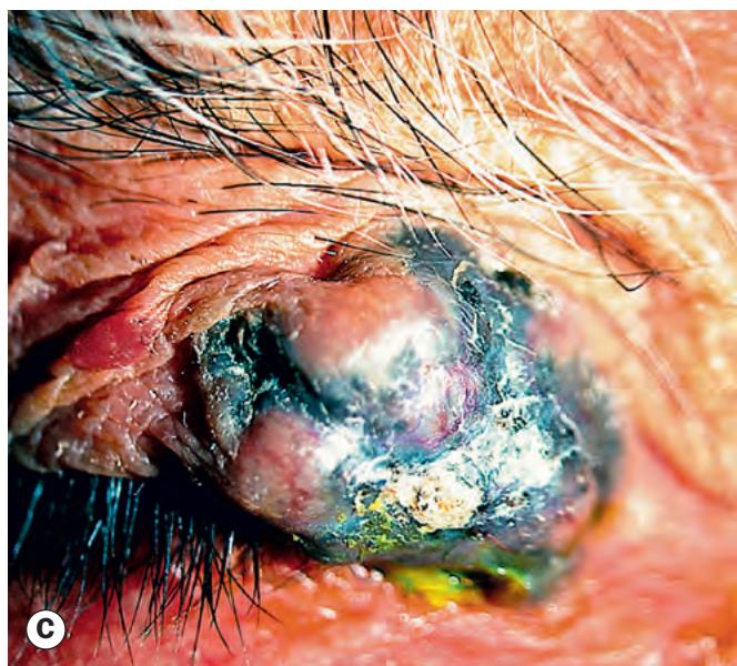
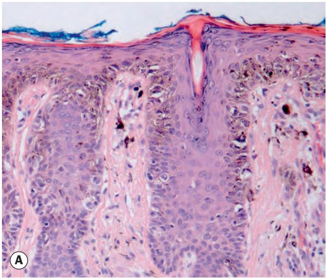
Fig. 1.25 Melanoma. (A) Histopathology shows melanoma cells within the dermis; (B) superficial spreading melanoma; (C) nodular melanoma
(Courtesy of L Horton – fig. A; S Delva – fig. C)
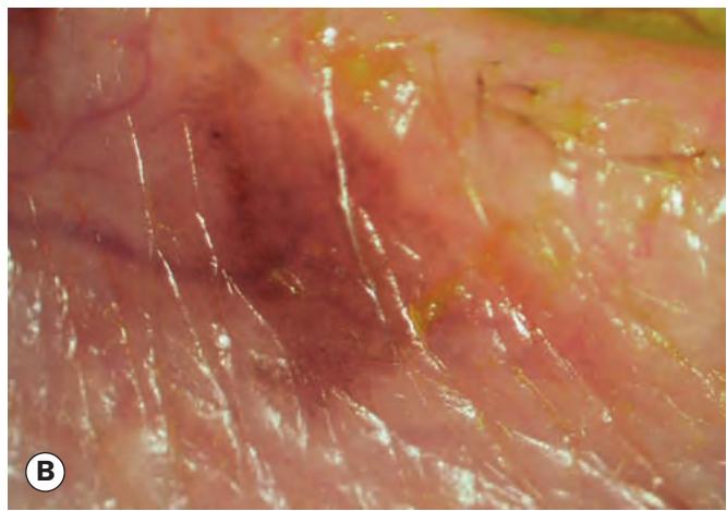
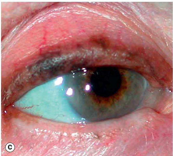
Fig. 1.24 Lentigo maligna of the eyelid. (A) Histopathology shows melanoma cells proliferating within the basal layers of the epidermis; (B) early lentigo maligna; (C) melanoma arising from lentigo maligna
(Courtesy of J Harry – fig. A)

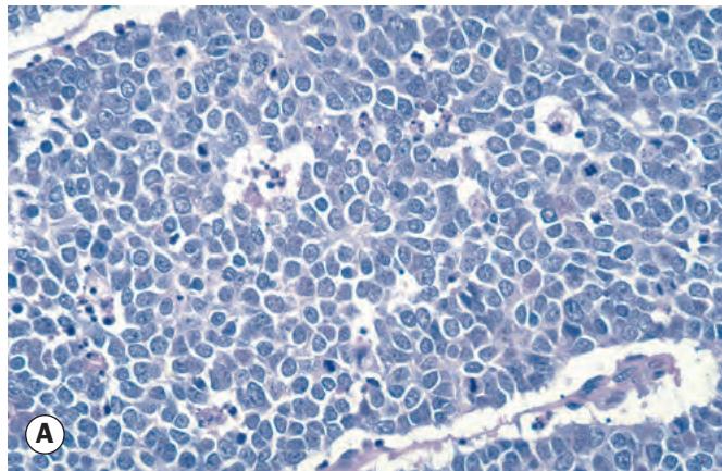
Fig. 1.26 Merkel cell carcinoma. (A) Histopathology shows a sheet of Merkel cells; (B) clinical appearance
(Courtesy of J Harry and G Misson, from Clinical Ophthalmic Pathology, Butterworth-Heinemann 2001 – fig. A)
Treatment of malignant tumours
Biopsy
Biopsy can be (i) incisional, using a blade or a biopsy punch, in which only part of the lesion is removed for histological diagnosis, or (ii) excisional, in which the entire lesion is removed; the latter may consist of shave excision using a blade to remove shallow epithelial tumours, such as papillomas and seborrhoeic keratosis, or full-thickness skin excision for tumours that are not confined to the epidermis.