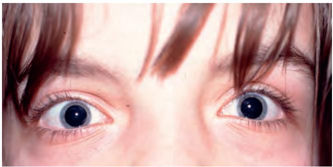
Fig. 1.80 Telecanthus
Blepharophimosis, ptosis and epicanthus inversus syndrome
Blepharophimosis, ptosis and epicanthus inversus syndrome (BPES) is a complex of eyelid malformations consisting of moderate to severe symmetrical ptosis with poor levator function, telecanthus, epicanthus inversus (see Fig. 1.79A), manifesting with small palpebral fissures (Fig. 1.81). Other minor facial anomalies are commonly present. Inheritance is usually autosomal dominant; both BPES type I (with premature ovarian failure) and BPES type II (without premature ovarian failure) are caused by mutations in the FOXL2 gene on chromosome 3. Treatment initially involves correction of epicanthus and telecanthus, followed later by bilateral frontalis suspension. It is also important to treat amblyopia, which is present in about 50%.
Epiblepharon
Epiblepharon comprises an extra horizontal fold of skin stretching across the anterior lid margin; it is very common in individuals of Eastern Asian ethnicity. The lashes are directed vertically, especially in the medial part of the lid (Figs 1.82A and B). When the fold of skin is pulled down the lashes turn out and the normal location of the lid becomes apparent (Fig. 1.82C); it should not be confused with the much less common congenital entropion (see next). Treatment is not required in the majority of Caucasians because spontaneous resolution with age is usual. Persistent cases may be treated surgically.
Congenital entropion
Upper lid entropion is usually secondary to the mechanical effects of microphthalmos, which cause variable degrees of upper lid inversion. Lower lid entropion (Fig. 1.83) is generally caused by maldevelopment of the inferior retractor aponeurosis. Treatment involves the excision of a strip of skin and muscle, and fixation of the skin crease to the tarsal plate (Hotz procedure).
Coloboma
A congenital coloboma is an uncommon, unilateral or bilateral, partial- or full-thickness eyelid defect. It occurs when eyelid development is incomplete, due to either failure of migration of lid ectoderm to fuse the lid folds or to mechanical forces such as amniotic bands. Colobomata elsewhere in the eye, as well as a range of other associations, may be present. The treatment of small defects involves primary closure, while large defects require skin grafts and rotation flaps.
Upper lid colobomas occur at the junction of the middle and inner thirds (Fig. 1.84A); relatively strong associations include cryptophthalmos (see below), facial abnormalities and Goldenhar syndrome.
Lower lid colobomas occur at the junction of the middle and outer thirds (Fig. 1.84B) and are frequently associated with systemic conditions.
Treacher Collins syndrome (mandibulofacial dysostosis) is a genetically heterogeneous condition characterized by malformation of derivatives of the first and second branchial arches, principally mandibular and ear anomalies. Lower eyelid coloboma is a feature; ocular anomalies also described include slanted palpebral apertures, cataract, microphthalmos and lacrimal atresia.
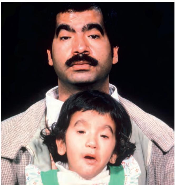
Fig. 1.81 Blepharophimosis ptosis and epicanthus inversus syndrome
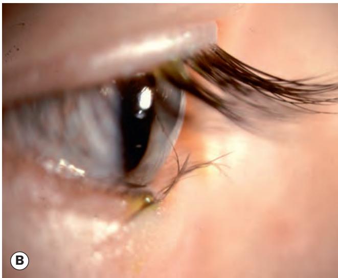
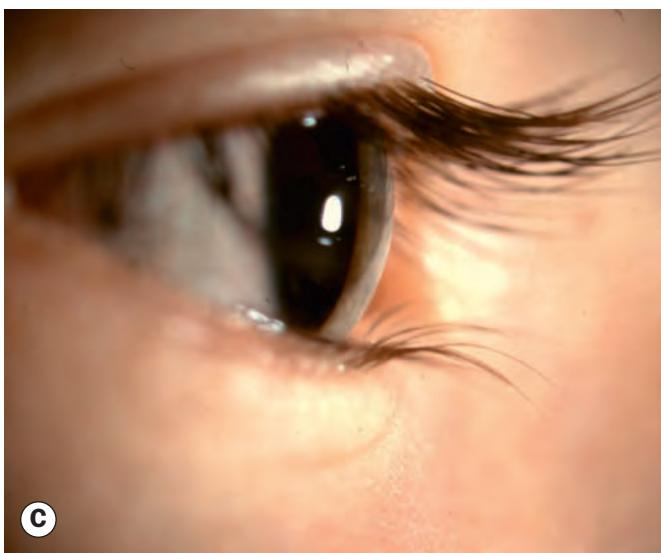
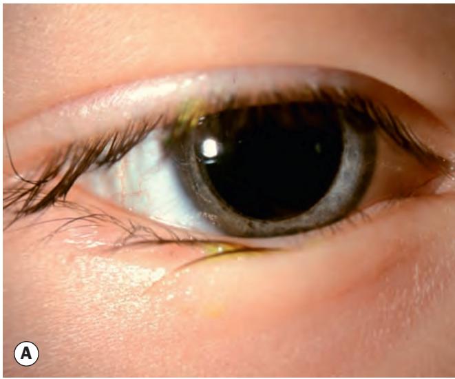
Fig. 1.82 (A) Epiblepharon; (B) lashes pointing upwards; (C) normal position of lashes following manual correction
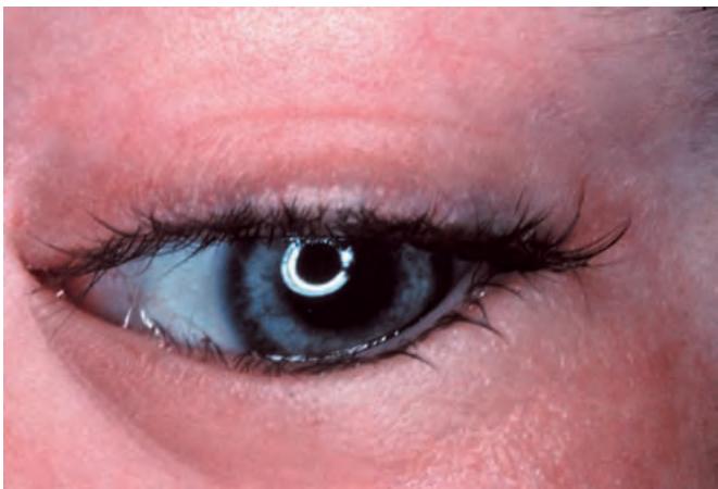
Fig. 1.83 Congenital lower lid entropion