Diagnosis
The upper eyelid is typically extremely lax, often with substantial excess loose upper lid skin (Fig. 1.72A). The tarsal plate has a rubbery consistency (Fig. 1.72B); the lid is very easy to evert (Fig. 1.72C), to fold and to pull away from the eye.
Papillary conjunctivitis of the superior tarsal conjunctiva may be intense (Fig. 1.72D).
Keratopathy. Punctate keratopathy, filamentary keratitis and superior superficial vascularization may be present.
Other findings may include eyelash ptosis, lacrimal gland prolapse, ectropion and aponeurotic ptosis. Patients with both FES and OSA seem to have a considerably higher than average prevalence of glaucoma.
Investigation for OSA should be considered in most cases of FES, particularly if the patient reports substantial snoring and/or excessive daytime sleepiness.
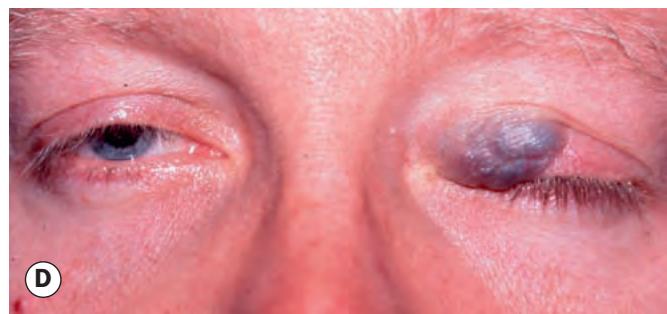
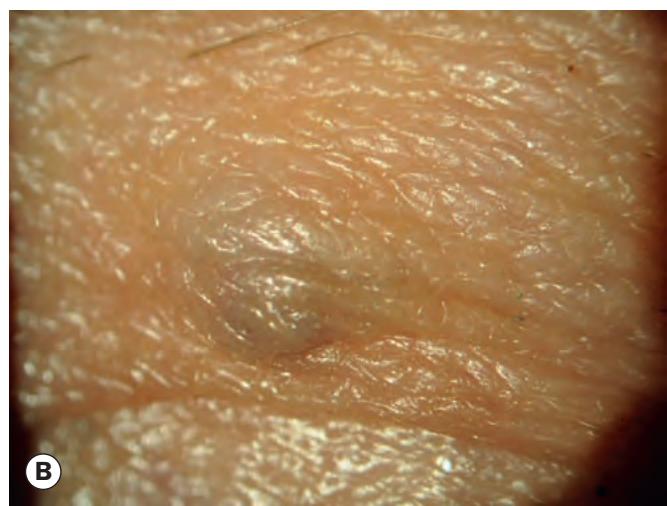
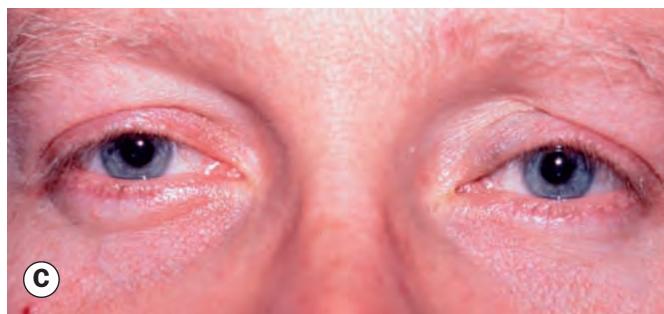

Fig. 1.71 Eyelid varices. (A) Typical appearance of a commonly seen small varix; (B) magnified view; (C) larger lesion, probably with orbital involvement, before Valsalva manoeuvre; (D) during Valsalva
(Courtesy of G Rose – figs C and D)
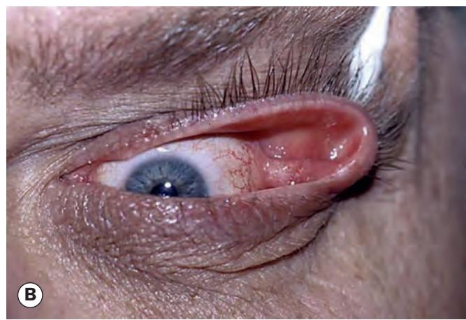
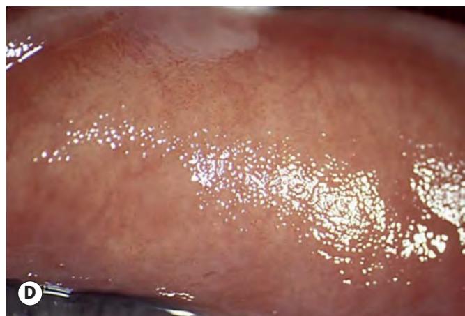
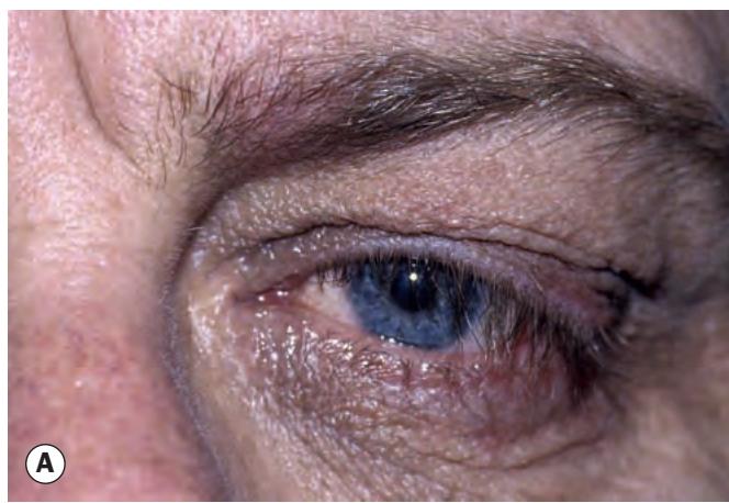
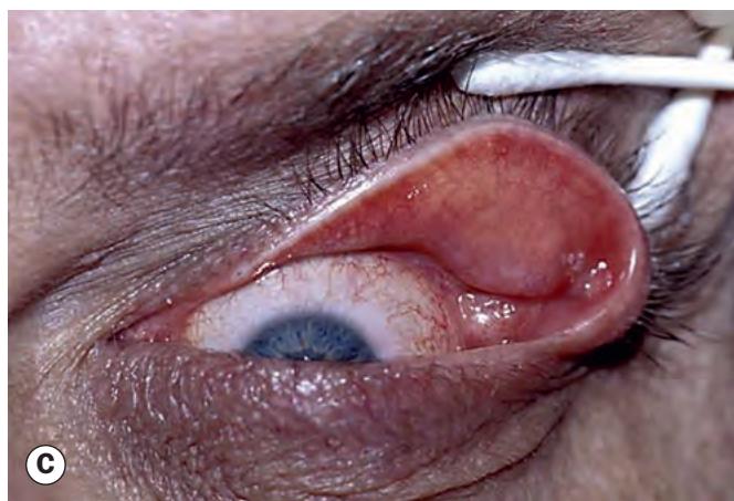
Fig. 1.72 Floppy eyelid syndrome. (A) Redundant upper lid skin; (B) loose and rubbery tarsal plates; (C) very easily everted eyelid; (D) superior tarsal papillary conjunctivitis