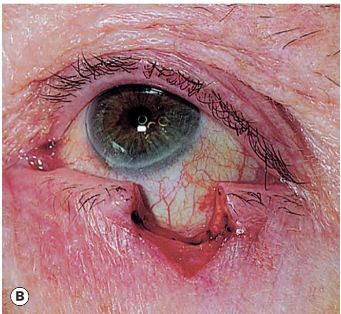

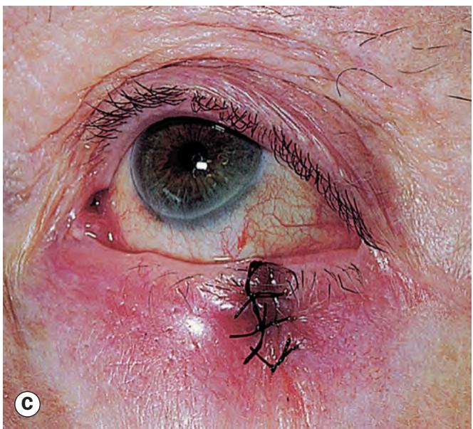
Fig. 1.63 Horizontal lid shortening to correct ectropion.
(A) Marking; (B) excision of a pentagon; (C) closure
(Courtesy of AG Tyers and JRO Collin, from Colour Atlas of Ophthalmic Plastic Surgery, Butterworth-Heinemann 2001)

Fig. 1.64 Cicatricial ectropion
(Courtesy of A Pearson)
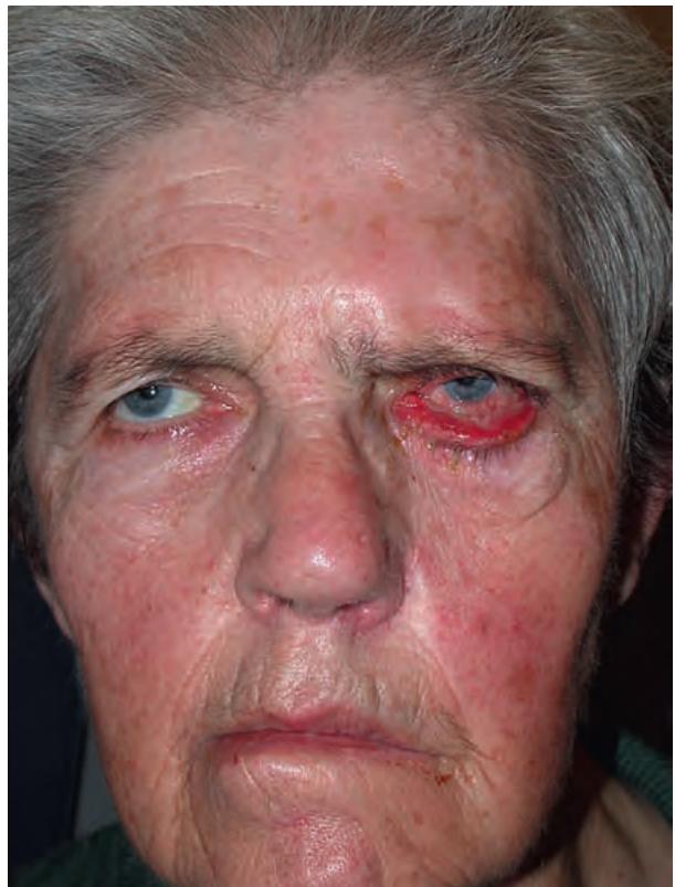
Fig. 1.65 Left facial palsy and severe paralytic ectropion
Entropion
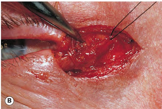
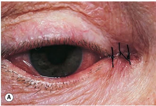
Fig. 1.66 Permanent treatment of paralytic ectropion.
(A) Medial canthoplasty; (B) lateral canthal sling – refashioned canthal tendon from the lower lid is passed through a buttonhole in the tendon from the upper lid
(Courtesy of AG Tyers and JRO Collin, from Colour Atlas of Ophthalmic Plastic Surgery, Butterworth-Heinemann 2001)
Mechanical ectropion
Mechanical ectropion is caused by tumours on or near the lid margin that mechanically evert the lid. Treatment involves removal of the cause if possible, and correction of significant horizontal lid laxity.
ENTROPION
Involutional entropion
Introduction
Involutional (age-related) entropion affects mainly the lower lid. The constant rubbing of the lashes on the cornea in long-standing entropion (pseudotrichiasis – Fig. 1.67A) may cause irritation, corneal punctate epithelial erosions and, in severe cases, pannus formation and ulceration. Aetiological factors include:
Horizontal lid laxity caused by stretching of the canthal tendons and tarsal plate.
Vertical lid instability caused by attenuation, dehiscence or disinsertion of the lower lid retractors. Weakness of the latter is recognized by decreased excursion of the lower lid in downgaze.
Over-riding of the pretarsal by the preseptal orbicularis during lid closure tends to move the lower border of the tarsal plate anteriorly, away from the globe, and the upper border towards the globe, thus tipping the lid inwards (Fig. 1.67B).
Orbital septum laxity with prolapse of orbital fat into the lower lid.