Cicatricial ectropion
Cicatricial ectropion is caused by scarring or contracture of the skin and underlying tissues, which pulls the eyelid away from the globe (Fig. 1.64). If the skin is pushed up over the orbital margin with a finger the ectropion will be relieved. Opening the mouth tends to accentuate the eversion. Depending on the cause, both lids may be involved and the defect may be local (e.g. trauma) or general (e.g. burns, dermatitis, ichthyosis). Mild localized cases are treated by excision of the offending scar tissue combined with a procedure that lengthens vertical skin deficiency, such as Z-plasty. Severe generalized cases require transposition flaps or free skin grafts; sources of skin include the upper lids, posterior auricular, preauricular and supraclavicular areas.
Paralytic ectropion/facial nerve palsy
Introduction
Paralytic ectropion is caused by ipsilateral facial nerve palsy (Fig. 1.65) and is associated with retraction of the upper and lower lids and brow ptosis; the latter may mimic narrowing of the palpebral aperture.
Complications include exposure keratopathy due to lagophthalmos, and watering caused by malposition of the inferior lacrimal punctum, failure of the lacrimal pump mechanism and an increase in tear production resulting from corneal exposure.
Treatment
Temporary measures may be instituted to protect the cornea in anticipation of spontaneous recovery of facial nerve function.
○ Lubrication with higher viscosity tear substitutes during the day, with instillation of ointment and taping shut of the lids during sleep, are usually adequate in mild cases.
○ Botulinum toxin injection into the levator to induce temporary ptosis.
☐ Temporary tarsorrhaphy may be necessary, particularly in patients with a poor Bell phenomenon with the cornea remaining exposed when the patient attempts to blink; the lateral aspects of the upper and lower lids are sutured together.
Permanent treatment should be considered when there is irreversible damage to the facial nerve as may occur following removal of an acoustic neuroma, or when no further improvement has occurred for 6-12 months in a Bell palsy.
☐ Medial canthoplasty may be performed if the medial canthal tendon is intact. The eyelids are sutured together medial to the lacrimal puncta (Fig. 1.66A) so that the puncta become inverted and the fissure between the inner canthus and puncta is shortened.
○ A lateral canthal sling or tarsal strip may be used to correct residual ectropion and raise the lateral canthus (Fig. 1.66B).
○ Upper eyelid lowering by levator disinsertion.
○ Gold weight implantation in the upper lid can assist closure.
○ A small lateral tarsorrhaphy is usually cosmetically acceptable.

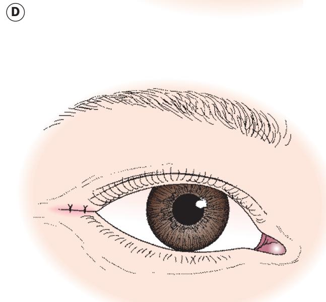
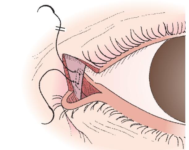


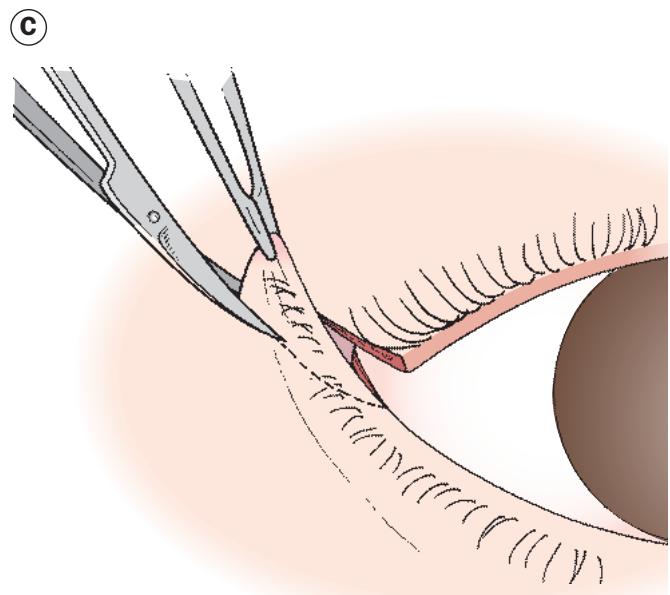


Fig. 1.62 Lateral tarsal strip procedure. (A) Lateral canthotomy; (B) cantholysis – the lower limb of the lateral canthal tendon is cut away from the inferior orbital rim; (C) the anterior and posterior lid lamellae are divided, and a ‘strip’ of tendon/ lateral tarsal plate dissected out; (D) the strip is shortened and then reattached to the inner aspect of the orbital rim periosteum with 4-0 absorbable suture; (E) excess lid margin is trimmed; (F) the skin incision (canthotomy) is closed (Courtesy of JA Nerad, from Techniques in Ophthalmic Plastic Surgery, Saunders 2010)