Conjunctiva–Müller resection
This involves excision of Müller muscle and overlying conjunctiva (Fig. 1.58A) with reattachment of the resected edges (Fig. 1.58B).
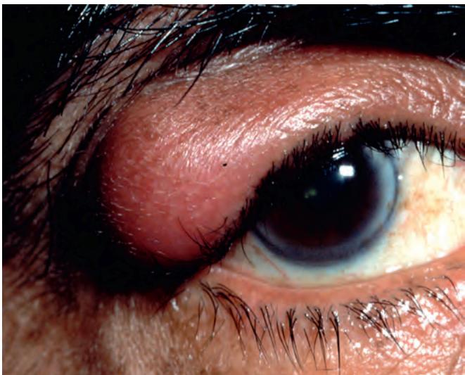
Fig. 1.56 Mechanical ptosis due to a neurofibroma
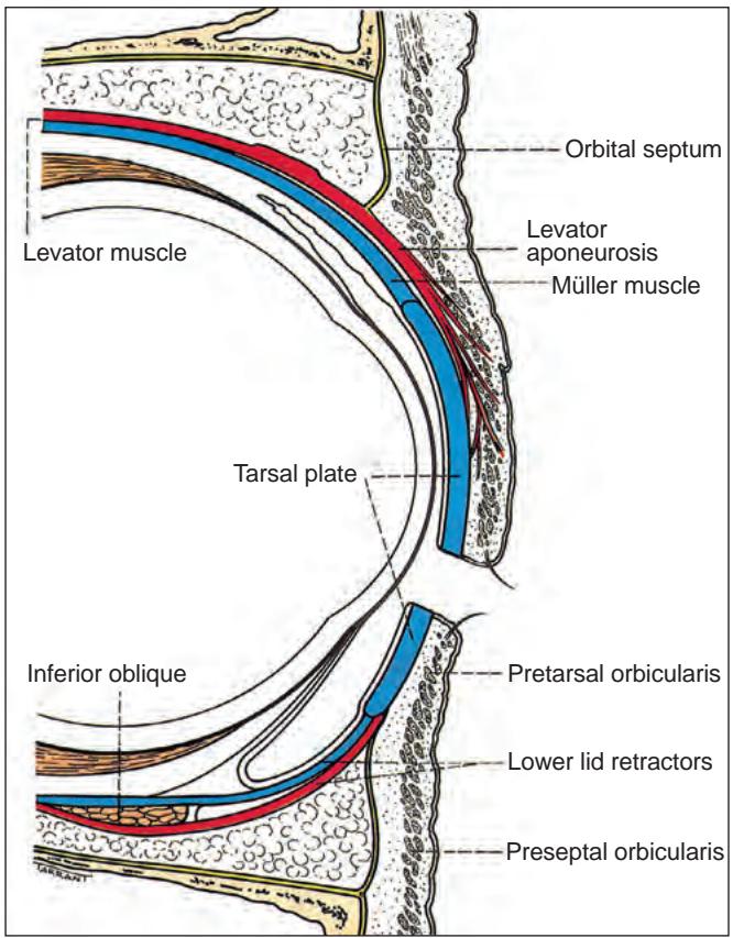
Fig. 1.57 Anatomy of the eyelid
The maximal elevation achievable is 2–3 mm, so it is used in cases of mild ptosis with good (at least 10 mm) levator function, which includes most cases of Horner syndrome and mild congenital ptosis.
Levator advancement (resection)
In this technique the levator complex is shortened through either an anterior – skin (Fig. 1.59) – or posterior – conjunctival – approach. Indications include ptosis of any cause, provided residual levator function is at least 5 mm. The extent of resection is determined by the severity of the ptosis and the amount of levator function.
Brow (frontalis) suspension
Brow (frontalis) suspension is used for severe ptosis (>4 mm) with very poor levator function (<4 mm) from a variety of causes, typical indications being ptosis associated with third nerve palsy, blepharophimosis syndrome and following an unsatisfactory result from previous levator resection. The tarsal plate is suspended from the frontalis muscle with a sling consisting of autologous fascia lata (Fig. 1.60) or non-absorbable material such as prolene or silicone.
ECTROPION
Involutional ectropion
Introduction
Involutional (age-related) ectropion affects the lower lid of elderly patients. It causes epiphora (tear overflow) and may exacerbate ocular surface disease. The red appearance of the exposed conjunctiva is cosmetically poor. In long-standing cases the tarsal conjunctiva may become chronically inflamed, thickened and keratinized (Fig. 1.61). Aetiological factors include:
Horizontal lid laxity can be demonstrated by pulling the central part of the lid 8 mm or more from the globe, with a failure to snap back to its normal position on release without the patient first blinking.
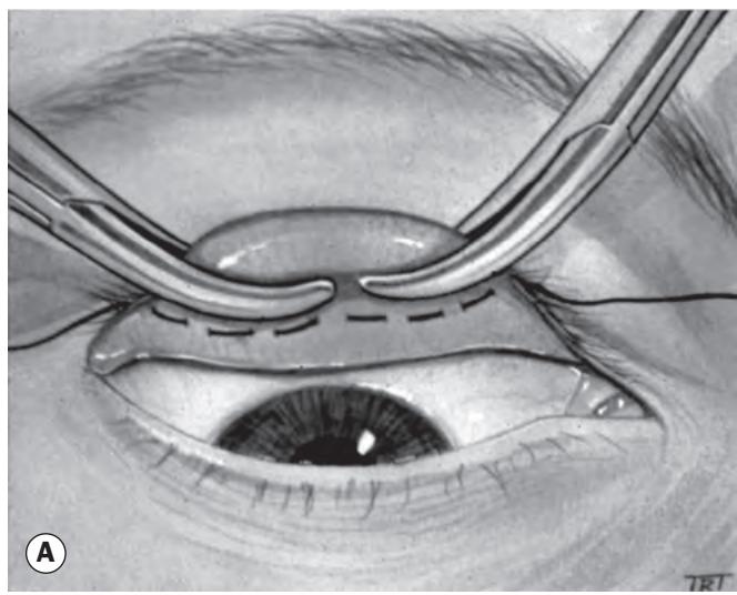
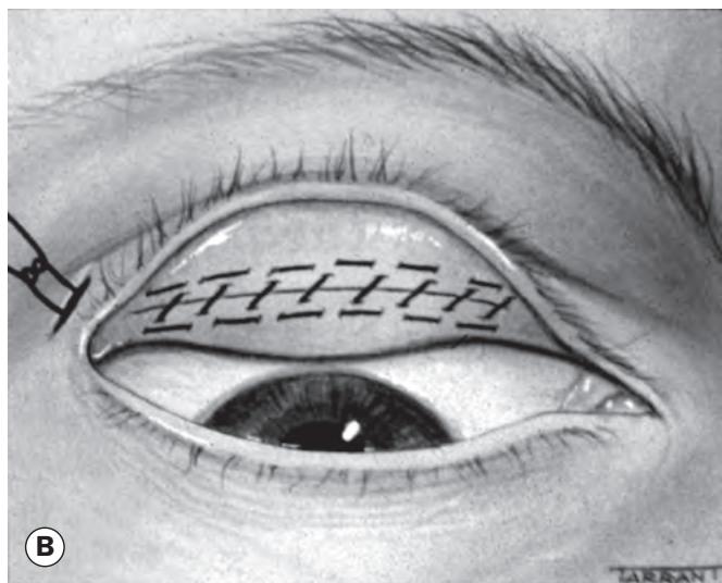
Fig. 1.58 Conjunctiva–Müller resection. (A) Clamping of conjunctiva and Müller muscle; (B) appearance after excision and suturing