Clinical evaluation
General
The age at onset of ptosis and its duration will usually distinguish congenital from acquired cases. If the history is ambiguous, old photographs may be helpful. It is also important to enquire about symptoms of possible underlying systemic disease, such as associated diplopia, variability of ptosis during the day and excessive fatigue.
Pseudoptosis
A false impression of ptosis may be caused by the following:
Lack of support of the lids by the globe may be due to an orbital volume deficit associated with an artificial eye, microphthalmos, phthisis bulbi (Fig. 1.47A), or enophthalmos.
Contralateral lid retraction, which is detected by comparing the levels of the upper lids, remembering that the margin of the upper lid normally covers the superior 2 mm of the cornea (Fig. 1.47B).
Ipsilateral hypotropia causes pseudoptosis because the upper lid follows the globe downwards (Fig. 1.47C). It disappears when the hypotropic eye assumes fixation on covering the normal eye.
Brow ptosis due to excessive skin on the brow, or seventh nerve palsy, which is diagnosed by manually elevating the eyebrow (Fig. 1.47D).
Dermatochalasis. Overhanging skin on the upper lids (Fig. 1.48) may be mistaken for ptosis, but may also cause mechanical ptosis.
Measurements
Margin–reflex distance is the distance between the upper lid margin and the corneal reflection of a pen torch held by the examiner on which the patient fixates (Fig. 1.49); the normal measurement is 4–5 mm.
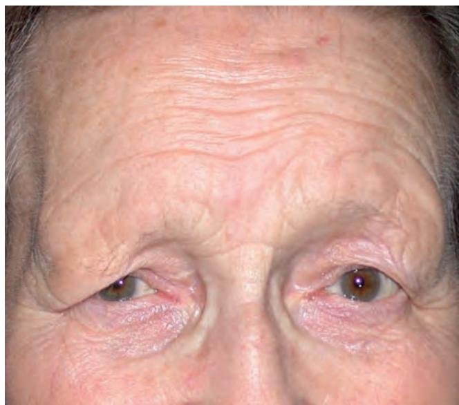
Fig. 1.48 Marked right dermatochalasis and brow ptosis (Courtesy of S Chen)

Fig. 1.49 Margin–reflex distance. (A) Normal; (B) mild ptosis; (C) moderate ptosis; (D) severe ptosis
Palpebral fissure height is the distance between the upper and lower lid margins, measured in the pupillary plane (Fig. 1.50). The upper lid margin normally rests about 2 mm below the upper limbus and the lower 1 mm above the lower limbus. This measurement is shorter in males (7–10 mm) than in females (8–12 mm). Unilateral ptosis can be quantified by comparison with the contralateral side. Ptosis may be graded as mild (up to 2 mm), moderate (3 mm) and severe (4 mm or more).

Fig. 1.51 Measurement of levator function
Levator function (upper lid excursion) is measured by placing a thumb firmly against the patient’s brow to negate the action of the frontalis muscle, with the eyes in downgaze (Fig. 1.51A). The patient then looks up as far as possible and the amount of excursion is measured with a rule (Fig. 1.51B). Levator function is graded as normal (15 mm or more), good (12–14 mm), fair (5–11 mm) and poor (4 mm or less).
Upper lid crease is taken as the vertical distance between the lid margin and the lid crease in downgaze. In females it measures about 10 mm and in males 8 mm. Absence of the crease in a patient with congenital ptosis is evidence of poor levator function, whereas a high crease suggests an aponeurotic defect (usually involutional). The skin crease is also used as a guide to the initial incision in some surgical procedures.
Pretarsal show is the distance between the lid margin and the skin fold with the eyes in the primary position.
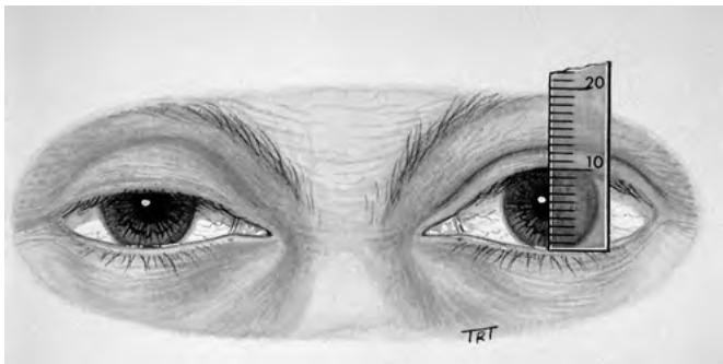
Fig. 1.50 Measurement of palpebral fissure height
Associated signs
The pupils should be examined to exclude Horner syndrome and a subtle pupil-involving third nerve palsy – the latter is an unlikely acute clinical presentation (see Ch. 19).
Increased innervation may flow to the levator muscle of a unilateral ptosis, particularly in upgaze. Associated increased innervation to the contralateral normal levator will result in lid retraction. The examiner should therefore manually elevate the ptotic lid and look for drooping of the opposite lid. If this occurs, the patient should be warned that surgical correction may induce a lower position in the opposite lid.
Fatigability is tested by asking the patient to look up without blinking for 30–60 seconds. Progressive drooping of one or both lids, or an inability to maintain upgaze, is suggestive of myasthenia gravis (see Ch. 19). Myasthenic ptosis may show an overshoot of the upper lid on saccade from downgaze to the primary position (Cogan twitch sign) and a ‘hop’ on side-gaze.
Ocular motility defects, particularly of the superior rectus, must be evaluated in patients with congenital ptosis. Correction of an ipsilateral hypotropia may improve the degree of ptosis. Deficits consistent with a subtle or partial third nerve paresis should be identified.
Jaw-winking can be identified by asking the patient to chew and move the jaws from side to side (see below).
The Bell phenomenon is tested by manually holding the lids open, asking the patient to try to shut the eyes and observing upward and outward rotation of the globe. A weak Bell phenomenon carries a variable risk of postoperative exposure keratopathy, particularly following large levator resections or suspension procedures.
The tear film should be inspected – a poor volume or unstable film may be worsened by ptosis surgery and should be addressed preoperatively as far as possible.