Tick infestation of the eyelid
Ticks (see Fig. 11.69B) can attach themselves to the eyelid and should be removed at the earliest opportunity in order to minimize the risk of contracting a tick-borne zoonosis such as Lyme disease, Rocky Mountain fever or tularaemia. If the tick is attached some distance from the eye such that spray can safely be applied, an insect repellent containing pyrethrin or a pyrethroid should be sprayed on the tick twice at intervals of a minute; alternatively a scabies cream containing permethrin can be applied. These have a toxic effect that prevents the tick from injecting saliva, and after 24 hours it should drop off or can be removed with fine-tipped forceps at the slit lamp (blunt-tipped needle-holders are an alternative in restrained small children). It is critical that the tick is detached as close to its skin attachment as possible in order to remove its head and mouthparts, following which it might be retained in sealed packaging to permit identification if necessary. In areas endemic for Lyme disease, some authorities suggest routine antibiotic prophylaxis with doxycycline (in the absence of contraindications) following a confirmed deer tick bite, but as a minimum patients should be told to seek medical advice urgently at the onset of suspicious symptoms, particularly erythema migrans, over the subsequent few weeks. Lyme disease transmission is thought to require attachment of the tick for at least 36 hours.
Angular blepharitis
The infection is usually caused by Moraxella lacunata or S. aureus although other bacteria, and rarely herpes simplex, have also been implicated. Red, scaly, macerated and fissured skin is seen at the lateral and/or medial canthi of one or both eyes (Fig. 1.46A). Skin chafing secondary to tear overflow, especially at the lateral canthus, can cause a similar clinical picture, and may also predispose to infection (Fig. 1.46B). Associated papillary and follicular conjunctivitis may occur. Treatment involves topical chloramphenicol, bacitracin or erythromycin.
Childhood blepharokeratoconjunctivitis
Childhood blepharokeratoconjunctivitis is a poorly defined condition that tends to be more severe in Asian and Middle Eastern populations. Presentation is usually at about 6 years of age with recurrent episodes of anterior or posterior blepharitis, sometimes associated with recurrent styes or chalazia. Constant eye rubbing and photophobia may lead to misdiagnosis as allergic eye disease. Conjunctival changes include diffuse hyperaemia, bulbar phlyctens and follicular or papillary hyperplasia.
Corneal changes include superficial punctate keratopathy, marginal keratitis, peripheral vascularization and axial subepithelial haze. Treatment is with lid hygiene and topical antibiotic ointment at bedtime. Topical low-dose steroids (prednisolone 0.1% or fluorometholone 0.1%) and erythromycin syrup 125 mg daily for 4–6 weeks may also be used.
PTOSIS
Classification
Ptosis is an abnormally low position of the upper lid; it may be congenital or acquired.
Neurogenic ptosis is caused by an innervational defect such as third nerve paresis and Horner syndrome (see Ch. 19).
Myogenic ptosis is caused by a myopathy of the levator muscle itself, or by impairment of transmission of impulses at the neuromuscular junction (neuromyopathic). Acquired myogenic ptosis occurs in myasthenia gravis, myotonic dystrophy and progressive external ophthalmoplegia (see Ch. 19).
Aponeurotic or involutional ptosis is caused by a defect in the levator aponeurosis.
Mechanical ptosis is caused by the gravitational effect of a mass or by scarring.


Fig. 1.46 (A) Angular blepharitis; (B) tear overflow from a lax and rounded lateral canthus

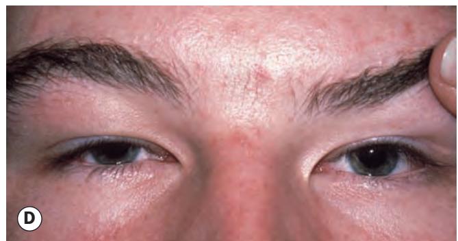
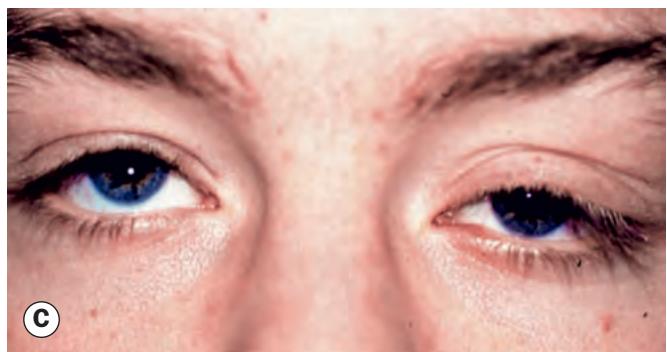
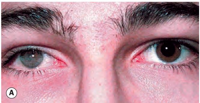
Fig. 1.47 Causes of pseudoptosis. (A) Right phthisis bulbi; (B) contralateral lid retraction; (C) ipsilateral hypotropia; (D) bilateral brow ptosis
(Courtesy of S Webber – figs C and D)