• Signs – staphylococcal blepharitis
○ Hard scales and crusting mainly located around the bases of the lashes; collarettes are cylindrical collections around lash bases (Fig. 1.42A).
○ Mild papillary conjunctivitis and chronic conjunctival hyperaemia are common.
○ Long-standing cases may develop scarring and notching (tylosis) of the lid margin, madarosis, trichiasis and poliosis.
☐ Associated tear film instability and dry eye syndrome are common.
○ Atopic keratoconjunctivitis may be present in patients with atopic dermatitis.
• Signs – seborrhoeic blepharitis
○ Hyperaemic and greasy anterior lid margins with soft scales and adherence of lashes to each other (Fig. 1.42B).
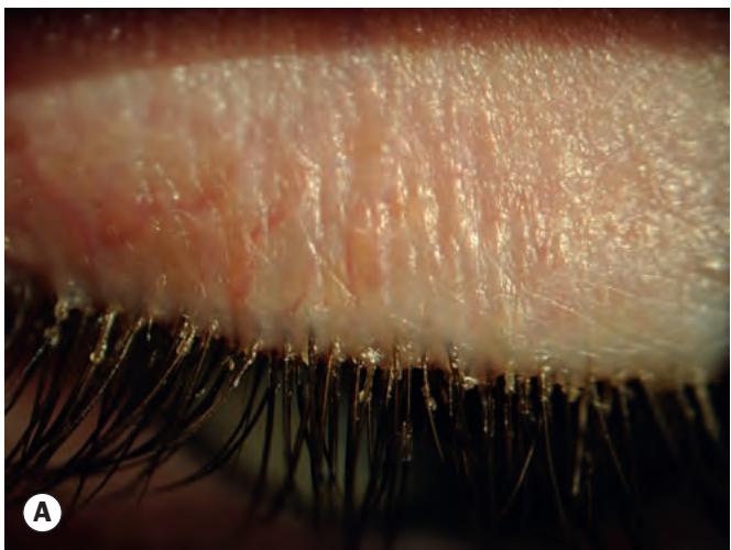

Fig. 1.42 Chronic anterior blepharitis. (A) Scales and crusting including collarettes in staphylococcal; (B) greasy lid margin with sticky lashes in seborrhoeic
Signs – posterior blepharitis (meibomian gland disease)
○ Excessive and abnormal meibomian gland secretion, manifesting as capping of meibomian gland orifices with oil globules (Fig. 1.43A).
○ Pouting, recession, or plugging of meibomian gland orifices (Fig. 1.43B).
○ Hyperaemia and telangiectasis of the posterior lid margin.
☐ Pressure on the lid margin results in expression of meibomian fluid that may be turbid or toothpaste-like (Fig. 1.43C); in severe cases the secretions become so inspissated that expression is impossible.
○ Lid transillumination may show gland loss and cystic dilatation of meibomian ducts.
☐ The tear film is oily and foamy and often unstable, and froth may accumulate on the lid margins (Fig. 1.43D) or inner canthi.
Demodex infestation may lead to cylindrical dandruff-like scaling (collarettes) around the base of eyelashes, though this is not always present. The mites can be demonstrated under ×16 slit lamp magnification by first manually clearing around the base of an eyelash then with fine forceps gently rotating the lash or moving it from side to side for 5–10 seconds, when if one or more mites (0.2–0.4 mm long – Fig. 1.44) does not emerge the lash should be gently epilated; slide microscopy can be performed on the mites or lashes if necessary.
Secondary changes include papillary conjunctivitis, inferior corneal punctate epithelial erosions, corneal scarring and vascularization including Salzmann nodular degeneration and advancing wave-like epitheliopathy-type changes, stye formation, marginal keratitis, and occasionally bacterial keratitis (especially in contact lens wearers) and phlyctenulosis.
Treatment
There is limited evidence to support any particular treatment protocol for blepharitis. Patients should be advised that a permanent cure is unlikely, but control of symptoms is usually possible. The treatment of anterior and posterior disease is broadly similar for both types, particular given that they commonly co-exist, but some treatments are fairly specific for one or the other.
Lid hygiene can be carried out once or twice daily initially; compliance and technique is highly variable.
○ A warm compress should first be applied for several minutes to soften crusts at the bases of the lashes.
○ Lid cleaning is subsequently performed to mechanically remove crusts and other debris, scrubbing the lid margins with a cotton bud or clean facecloth dipped in a warm dilute solution of baby shampoo or sodium bicarbonate.
☐ Commercially produced soap/alcohol impregnated pads for lid scrubbing are available and are often highly effective, but care should be taken not to induce mechanical irritation.
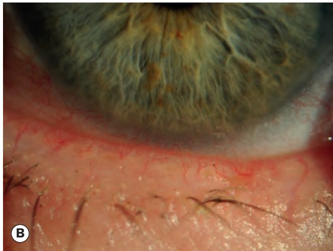

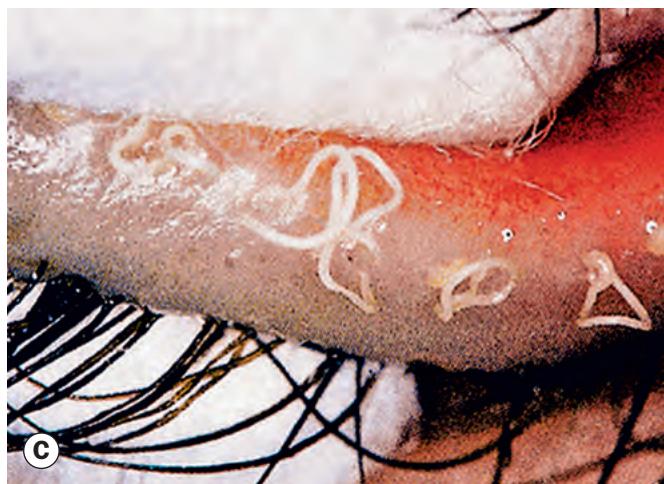
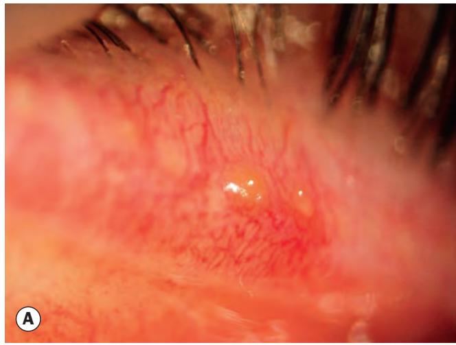
Fig. 1.43 Chronic posterior blepharitis. (A) Capping of meibomian gland orifices by oil globules; (B) hyperaemic, telangiectatic lid margin; (C) expressed toothpaste-like material; (D) froth on the eyelid margin
(Courtesy of J Silbert, from Anterior Segment Complications of Contact Lens Wear, Butterworth-Heinemann 1999 – fig. C)