Necrotizing fasciitis
Necrotizing fasciitis is a rare but commonly very severe infection involving subcutaneous soft tissue and the skin, with associated rapidly progressive necrosis. It is usually caused by S. pyogenes and occasionally S. aureus. The most frequent sites of involvement are the extremities, trunk and perineum, as well as postoperative wound sites. Unless early aggressive treatment is instituted, in the form of surgical debridement and high-dose intravenous antibiotics, death may result. Periocular infection is rare; redness and oedema are followed by the formation of large bullae and black discoloration of the skin due to necrosis (Fig. 1.38D).
VIRAL INFECTIONS
Molluscum contagiosum
Introduction
Molluscum contagiosum is a skin infection caused by a human-specific double-stranded DNA poxvirus that typically affects otherwise healthy children, with a peak incidence between 2 and 4 years of age. Transmission is by contact and subsequently by autoinoculation. Multiple, and occasionally confluent, lesions may develop in immunocompromised patients. Histopathology shows a central pit and lobules of hyperplastic epidermis with intracytoplasmic (Henderson–Patterson) inclusion bodies that displace the nuclear remnant to the edge of the cell. The bodies are small and eosinophilic near the surface, and large and basophilic deeper down (Fig. 1.39A).
Diagnosis
Single or multiple pale, waxy, umbilicated nodules develop (Fig. 1.39B); white cheesy material consisting of infected degenerate cells can be expressed from the lesion. Lesions on the lid margin (Fig. 1.39C) may shed virus into the tear film and give rise to a secondary ipsilateral chronic follicular conjunctivitis. Unless the lid margin is examined carefully the causative molluscum lesion may be overlooked.
Treatment
Spontaneous resolution will usually occur within a few months so treatment may not be necessary, particularly in children, unless complications such as a significant secondary conjunctivitis are problematic. Options include shave excision, cauterization, chemical ablation, cryotherapy and pulsed dye laser.
Herpes zoster ophthalmicus
Herpes zoster ophthalmicus (HZO – Fig. 1.40) is a common, generally unilateral infection caused by varicella-zoster virus. It is discussed in detail in Ch. 6.

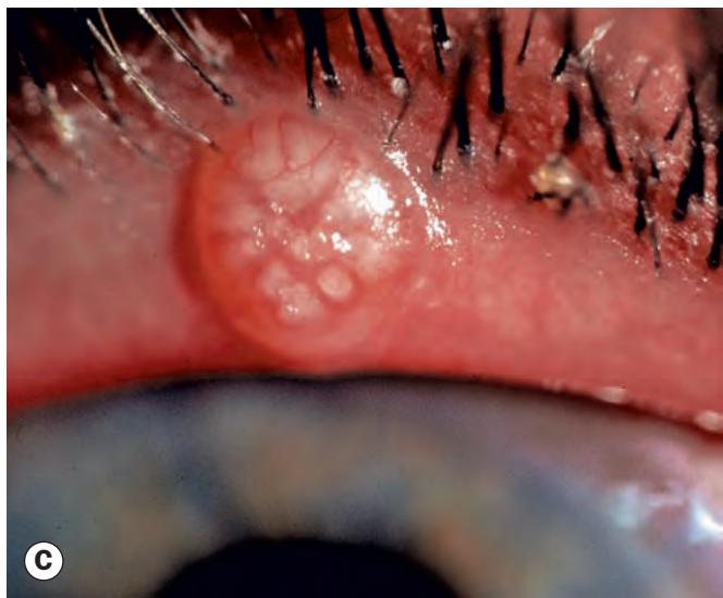

Fig. 1.39 Molluscum contagiosum. (A) Histopathology shows lobules of hyperplastic epidermis and a pit containing intracytoplasmic inclusion bodies; (B) multiple molluscum nodules; (C) lid margin nodule (Courtesy of A Garner – fig. A; N Rogers – fig. B)
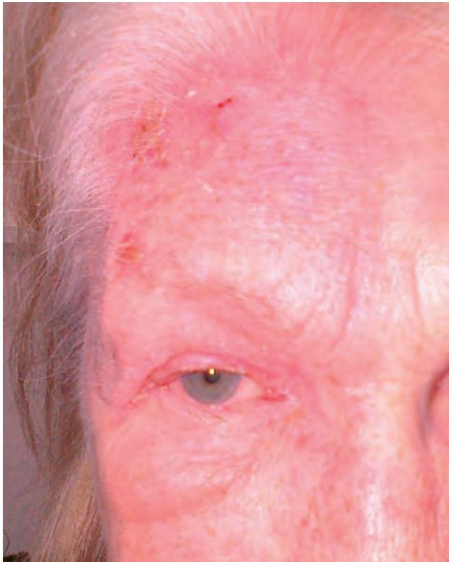
Fig. 1.40 Herpes zoster ophthalmicus – maculopapular crusting rash with periocular oedema, showing clear midline delineation
Herpes simplex
Introduction
Herpes simplex skin rash results from either primary infection or reactivation of herpes simplex virus previously dormant in the trigeminal ganglion. Prodromal facial and lid tingling lasting about 24 hours is followed by the development of eyelid and periocular skin vesicles (Fig. 1.41A) that break down over 48 hours (Fig. 1.41B). Although typically still confined to a single dermatome and with individual lesions that are often similar in appearance, the distribution of the herpes simplex skin rash contrasts with the sharply delineated unilateral involvement in HZO (see Fig. 1.40). There is commonly associated papillary conjunctivitis, discharge and lid swelling; dendritic corneal ulcers can develop, especially in atopic patients, in whom skin involvement can be extensive and very severe (eczema herpeticum – Fig. 1.41C).