• Relative contraindications
☐ Medial canthal lesions due to the high probability of lacrimal canalicular damage.
○ Upper eyelid tumours – conjunctival keratinization is common and difficult to manage.
○ Aggressive tumours such as SGC are relatively radioresistant, but higher-dose treatment may be effective.
Complications. Many of these can be minimized by appropriate shielding.
○ Skin damage and madarosis (eyelash loss).
○ Nasolacrimal duct stenosis following irradiation to the medial canthal area.
○ Conjunctival keratinization, dry eye, keratopathy and cataract.
○ Retinopathy and optic neuropathy.
Cryotherapy
Cryotherapy may be considered for small superficial BCCs; it can be a useful adjunct to surgery in some patients. Complications include skin depigmentation, madarosis and conjunctival overgrowth.
DISORDERS OF THE EYELASHES
Misdirected lashes
Introduction
The roots of the eyelashes (cilia) lie against the anterior surface of the tarsal plate. The cilia pass between the main part of the orbicularis oculi and its more superficial part (Riolan muscle), exiting the skin at the anterior lid margin and curving away from the globe. It is particularly important to be familiar with the normal anatomical appearance of the lid margin in order to be able to identify the cause of eyelash misdirection. From anterior to posterior:
Eyelashes (cilia).
The grey line, by definition the border between the anterior (lashes, skin and orbicularis) and posterior (tarsal plate and conjunctiva) lamellae.
The meibomian gland orifices are located just anterior to the mucocutaneous junction. The edge of the tarsal plate is deep to the gland orifices; the glands themselves run vertically within the plate.
The mucocutaneous junction is where keratinized epithelium of the skin merges with conjunctival mucous membrane.
Conjunctiva lines the posterior margin of the lid.
Clinical features
Trauma to the corneal epithelium may cause punctate epithelial erosions, with ocular irritation often worsened by blinking. Corneal ulceration and pannus formation may occur in severe cases. The clinical appearance varies with the cause.
Trichiasis refers to misdirection of growth from individual follicles (Fig. 1.32A), rather than a more extensive inversion of the lid or lid margin. The follicles are at anatomically normal sites. It is commonly due to inflammation such as chronic blepharitis or herpes zoster ophthalmicus, but can also be caused by trauma, including surgery such as incision and curettage of a chalazion (Fig. 1.32B).
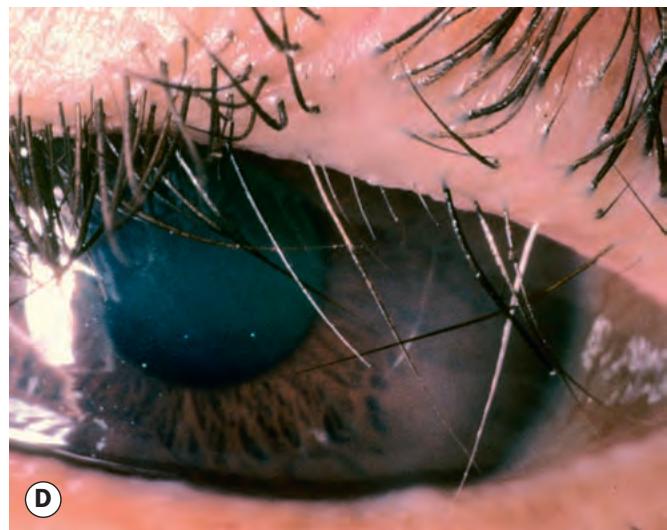
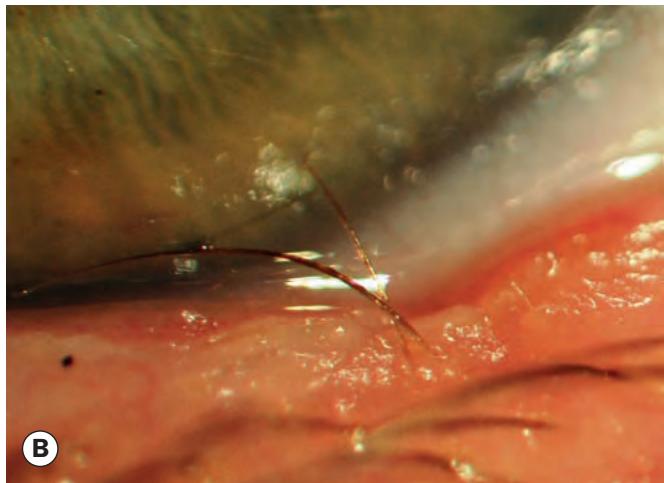

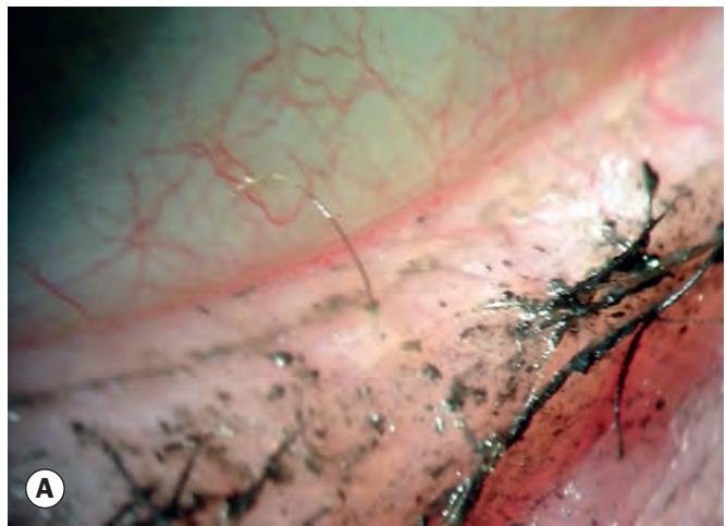
Fig. 1.32 Misdirected lashes. (A) Single trichiatic lash; (B) trichiasis associated with a lid notch following chalazion surgery; (C) marginal entropion showing rows of misdirected lashes, anterior migration of the mucocutaneous junction, and a rounded posterior lid margin; (D) acquired distichiasis
(Courtesy of S Chen – fig. A; R Bates – fig. D)
Marginal entropion has increasingly been recognized as a very common cause of eyelash misdirection, the mechanism of which is thought to be subtle cicatricial posterior lamellar shortening that rotates a segment of the lid margin towards the eye. The mucocutaneous junction migrates anteriorly and the posterior lid margin becomes rounded rather than physiologically square. Typically, numerous aligned lashes are involved (Fig. 1.32C).
Congenital distichiasis is a rare condition that occurs when a primary epithelial germ cell destined to differentiate into a meibomian gland develops instead into a complete pilosebaceous unit. The condition is frequently inherited in an autosomal dominant manner with high penetrance but variable expressivity. The majority of patients also manifest primary lymphoedema of the legs (lymphoedema– distichiasis syndrome). A partial or complete second row of lashes is seen to emerge at or slightly behind the meibomian gland orifices. The aberrant lashes tend to be thinner and shorter than normal cilia and are often directed posteriorly. They are usually well tolerated during infancy and may not become symptomatic until the age of about 5 years.
Acquired distichiasis is caused by metaplasia of the meibomian glands into hair follicles such that a variable number of lashes grow from meibomian gland openings. The most important cause is intense conjunctival inflammation (e.g. chemical injury, Stevens–Johnson syndrome, ocular cicatricial pemphigoid). In contrast to congenital distichiasis, the cilia tend to be non-pigmented and stunted (Fig. 1.32D), and are usually symptomatic.
Epiblepharon – see later.
Entropion. In contrast to marginal entropion, profound inversion of a substantial width of the lid is readily identified – see later.