Sebaceous gland carcinoma
Introduction
Sebaceous gland carcinoma (SGC) is a very rare, slowly growing tumour that most frequently affects the elderly, with a predisposition for females. It usually arises from the meibomian glands, although on occasion it may arise from the glands of Zeis or elsewhere. The tumour consists histopathologically of lobules of cells with pale foamy vacuolated lipid-containing cytoplasm and large hyperchromatic nuclei (Fig. 1.23A). Pagetoid spread refers to extension of a tumour within the epithelium, and is not uncommon. Overall mortality is 5–10%; adverse prognostic features include upper lid involvement, tumour size of 10 mm or more and duration of symptoms of more than 6 months.
Clinical features
In contrast to BCC and SCC, SGC occurs more commonly on the upper eyelid where meibomian glands are more numerous; there may be simultaneous involvement of both lids on one side (5%).
Yellowish material within the tumour is highly suggestive of SGC.
Nodular SGC presents as a discrete, hard nodule, most commonly within the upper tarsal plate (Fig. 1.23B), and may exhibit yellow discoloration due to the presence of lipid; it can be mistaken for a chalazion.
Spreading SGC infiltrates into the dermis and causes a diffuse thickening of the lid margin (Fig. 1.23C) often with eyelash distortion and loss, and can be mistaken for blepharitis.
Lentigo maligna and melanoma
Introduction
Melanoma rarely develops on the eyelids but is potentially lethal. Although pigmentation is a hallmark of skin melanomas, half of lid melanomas are non-pigmented and this may give rise to diagnostic difficulty. Features suggestive of melanoma include recent onset of a pigmented lesion, change in an existing pigmented lesion, irregular margins, asymmetrical shape, colour change or presence of multiple colours, and diameter greater than 6 mm.

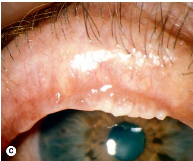
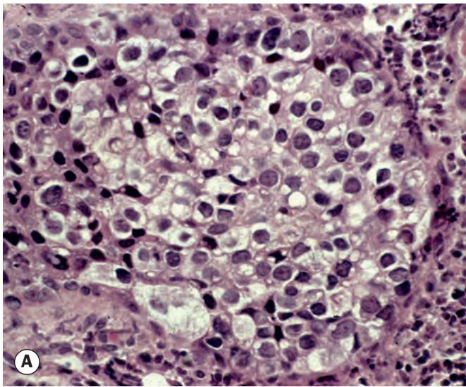
Fig. 1.23 Sebaceous gland carcinoma. (A) Histopathology shows cells with large hyperchromatic nuclei and vacuolated cytoplasm; (B) nodular tumour; (C) spreading tumour
(Courtesy of A Garner – fig. A; A Singh, from Clinical Ophthalmic Oncology, Saunders 2007 – fig. B; S Tuft – fig. C)
Lentigo maligna
Lentigo maligna (melanoma in situ, intraepidermal melanoma, Hutchinson freckle) is an uncommon condition that develops in sun-damaged skin in elderly individuals. Malignant change may occur, with infiltration of the dermis. Histopathology shows intraepidermal proliferation of spindle-shaped atypical melanocytes replacing the basal layer of the epidermis (Fig. 1.24A). Clinically lentigo maligna presents as a slowly expanding pigmented macule with an irregular border (Fig. 1.24B). Treatment is usually by excision. Nodular thickening and areas of irregular pig mentation are highly suggestive of malignant transformation (Fig. 1.24C).
Melanoma
Histopathology shows large atypical melanocytes invading the dermis (Fig. 1.25A). Superficial spreading melanoma is characterized by a plaque with an irregular outline and variable pigmentation (Fig. 1.25B). Nodular melanoma is typically a blue – black nodule surrounded by normal skin (Fig. 1.25C). Treatment is usually by wide excision and may include local lymph node removal. Radiotherapy, chemotherapy, biological and ‘targeted’ therapy may also be used, generally as adjuvants.
Merkel cell carcinoma
Merkel cells are a form of sensory receptor concerned with light touch. Merkel cell carcinoma is a rapidly growing, highly malignant tumour that typically affects older adults. Its rarity may lead to difficulty in diagnosis and delay in treatment, and 50% of patients have metastatic spread by presentation. A violaceous, well-demarcated nodule with intact overlying skin is seen, most frequently involving the upper eyelid (Fig. 1.26). Treatment is by excision, often with adjuvant therapy.
Kaposi sarcoma
Kaposi sarcoma is a vascular tumour that typically affects patients with AIDS. Many patients have advanced systemic disease although in some instances the tumour may be the only clinical manifestation of human immunodeficiency virus (HIV) infection. Histopathology shows proliferating spindle cells, vascular channels and inflammatory cells within the dermis (Fig. 1.27A). Clinically a pink, red-violet to brown lesion (Fig. 1.27B) develops, which may be mistaken for a haematoma or naevus. Treatment is by radiotherapy or excision, and by optimal control of AIDS where relevant.