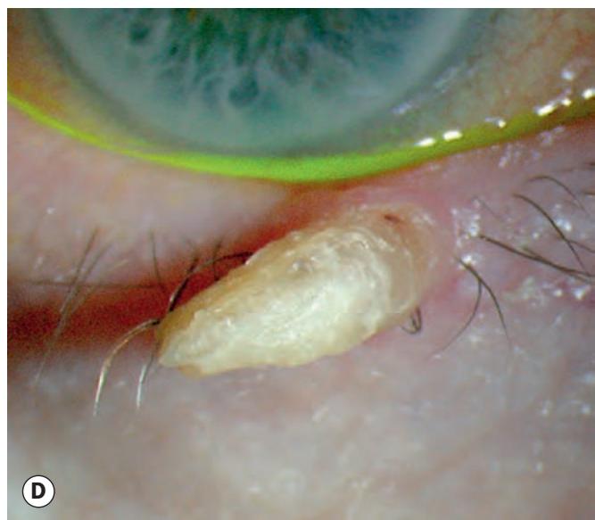
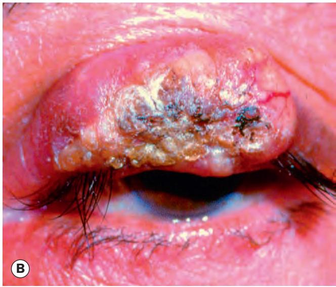
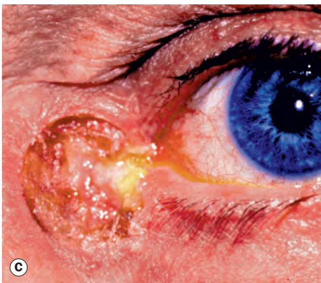

Fig. 1.21 Squamous cell carcinoma. (A) Histopathology shows acanthotic squamous epithelium and eosinophilic (pink) islands of dysplastic squamous epithelium within the dermis; (B) nodular tumour with surface keratosis; (C) ulcerating tumour; (D) cutaneous horn
(Courtesy of L Horton – fig. A; A Singh, from Clinical Ophthalmic Oncology, Saunders 2007 – fig. B; H Frank – fig. C; S Farley, T Cole and L Rimmer – fig. D)
Keratoacanthoma
Introduction
Keratoacanthoma is a rare, rapidly growing but subsequently regressing tumour that usually occurs in fair-skinned individuals with a history of chronic sun exposure. Immunosuppressive therapy is also a predisposing factor. It is regarded as falling within the spectrum of SCC, and although invasion and metastasis are rare, definitive treatment is usually indicated. Histopathologically, irregular thickened epidermis is surrounded by acanthotic squamous epithelium; a sharp transition from the thickened involved area to normal adjacent epidermis is referred to as shoulder formation (Fig. 1.22A); a keratin-filled crater may be seen.
Diagnosis
A pink dome-shaped hyperkeratotic lesion develops, often on the lower lid (Fig. 1.22B), and may double or treble in size within weeks (Fig. 1.22C). Growth then ceases for 2–3 months, after which spontaneous involution occurs, when a keratin-filled crater may develop (Fig. 1.22D). Complete involution may take up to a year and usually leaves an unsightly scar.
Treatment
Treatment generally involves complete surgical excision with a margin of at least 3 mm, or utilizing Mohs surgery; radiotherapy, cryotherapy or local chemotherapy are sometimes used. Observation is now regarded as inappropriate.
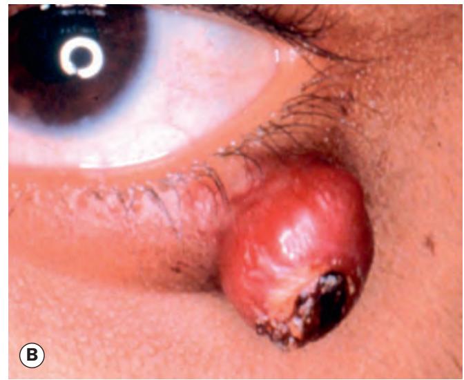

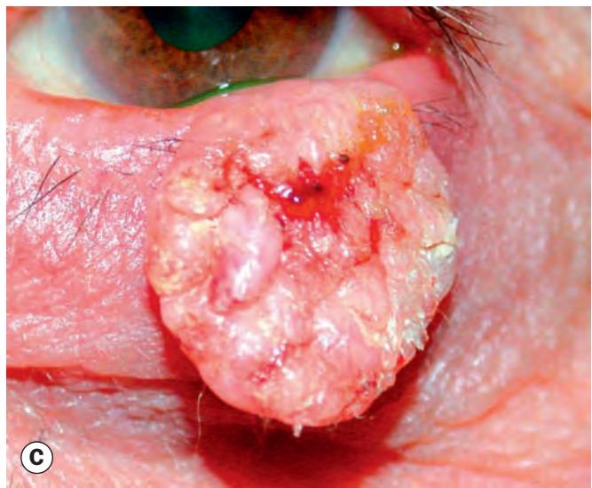
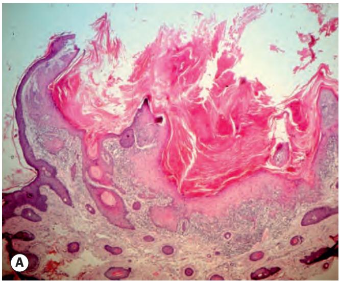
Fig. 1.22 Keratoacanthoma. (A) Histopathology shows irregularly thickened eosinophilic epidermis with a keratin-containing cup and well-marked shoulder formation; (B) hyperkeratotic nodule; (C) large tumour; (D) keratin-filled crater during involution