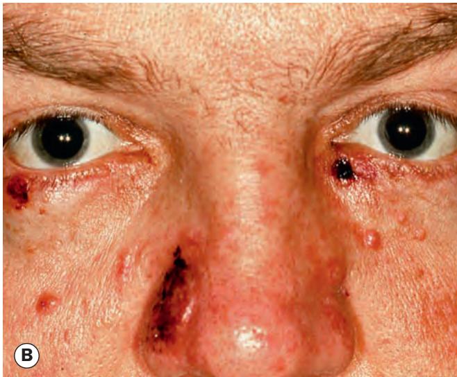
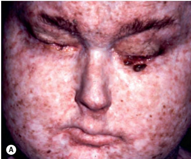
Fig. 1.17 Predispositions to eyelid malignancies. (A) Xeroderma pigmentosum; (B) Gorlin–Goltz syndrome
(Courtesy of J Krachmer, M Mannis and E Holland, from Cornea, Mosby 2005 – fig. B) and melanoma, which are commonly multiple. Conjunctival malignancies have also been reported.
Gorlin–Goltz syndrome (naevoid basal cell carcinoma syndrome) is a rare autosomal dominant (AD) disorder characterized by extensive congenital deformities of the eye, face, bone and central nervous system. Many patients develop multiple small BCC during the second decade of life (Fig. 1.17B) and are also predisposed to medulloblastoma, breast carcinoma and Hodgkin lymphoma.
Muir–Torre syndrome is a rare AD condition that predisposes to cutaneous and internal malignancies. Cutaneous tumours include BCC, sebaceous gland carcinoma and keratoacanthoma. Colorectal and genitourinary carcinomas are the most common systemic tumours.
Bazex syndrome can be used to describe two distinct conditions: (i) Bazex–Dupré–Christol syndrome, an
X-linked dominant condition characterized by multiple BCCs, commonly facial including the eyelids, associated with skin changes including follicular indentations without hairs on extensor surfaces (follicular atrophoderma), hypohidrosis and hypotrichosis; (ii) acrokeratosis paraneoplastica of Bazex, in which eczema-like and psoriatiform lesions are associated with an underlying malignancy of the upper respiratory or digestive tract.
Other predispositions include immunosuppression, prior retinoblastoma and albinism.
Basal cell carcinoma
Introduction
BCC is the most common human malignancy and typically affects older age groups. The most important risk factors are fair skin, inability to tan and chronic exposure to sunlight. Ninety per cent of cases occur in the head and neck and about 10% of these involve the eyelid. BCC is by far the most common malignant eyelid tumour, accounting for 90% of all cases. It most frequently arises from the lower eyelid, followed in relative frequency by the medial canthus, upper eyelid and lateral canthus. The tumour is slowly growing and locally invasive but non-metastasizing. Tumours located near the medial canthus are more prone to invade the orbit and sinuses, are more difficult to manage than those arising elsewhere and carry the greatest risk of recurrence. Tumours that recur following incomplete treatment tend to be more aggressive.
Histopathology
The tumour arises from the cells that form the basal layer of the epidermis. The cells proliferate downwards (Fig. 1.18A) and characteristically exhibit palisading at the periphery of a tumour lobule of cells (Fig. 1.18B). Squamous differentiation with the production of keratin results in a hyperkeratotic type of BCC. There can also be sebaceous and adenoid differentiation while the growth of elongated strands and islands of cells embedded in a dense fibrous stroma results in a sclerosing (morphoeic) type of tumour.
Clinical features
Eyelid BCC generally conforms to one of the morphological patterns below.
Nodular BCC is a shiny, firm, pearly nodule with small overlying dilated blood vessels. Initially, growth is slow and it may take the tumour 1–2 years to reach a diameter of 0.5 cm (Figs 1.19A and B).
Noduloulcerative BCC (rodent ulcer) is centrally ulcerated with pearly raised rolled edges and dilated and irregular blood vessels (telangiectasis) over its lateral margins (Fig. 1.19C); with time it may erode a large portion of the eyelid (Fig. 1.19D).
Sclerosing (morphoeic) BCC is less common and may be difficult to diagnose because it infiltrates laterally beneath
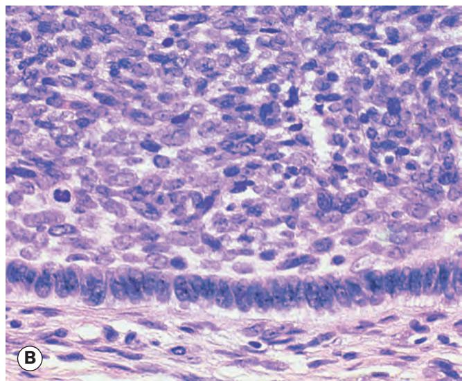

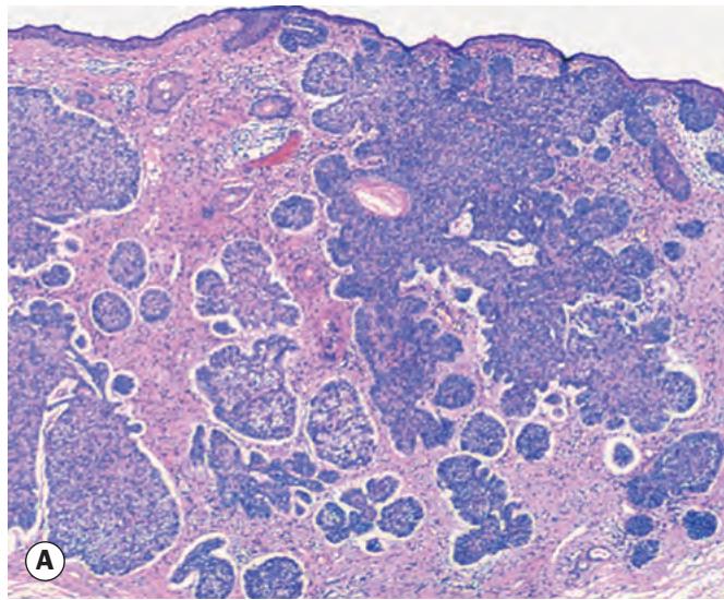
Fig. 1.18 Histopathology of basal cell carcinoma. (A) Histopathology shows downward proliferation of lobules of basophilic (purple) cells; (B) palisading of cells at the periphery of a tumour lobule (Courtesy of J Harry)