Sturge–Weber syndrome
Sturge–Weber syndrome (encephalotrigeminal angiomatosis) is a congenital, sporadic phacomatosis.
Port-wine stain, extending over the area corresponding to the distribution of one or more branches of the trigeminal nerve.
Leptomeningeal haemangioma involving the ipsilateral parietal or occipital region may cause contralateral focal or generalized seizures, hemiparesis or hemianopia.
Ocular features may include ipsilateral glaucoma, episcleral haemangioma, iris heterochromia and diffuse choroidal haemangioma (see Ch. 12).
Pyogenic granuloma
Pyogenic granuloma is a rapidly growing vascularized proliferation of granulation tissue that is usually antedated by surgery, trauma or infection, although some cases are idiopathic. Clinically there is a painful, rapidly growing, vascular granulating polypoidal lesion (Fig. 1.15) that may bleed following relatively trivial trauma. Treatment of cutaneous lesions involves excision; conjunctival pyogenic granuloma is discussed in Ch. 5.
Neurofibroma
Cutaneous neurofibromas are benign nerve tumours, usually nodular or pedunculated, that can be found anywhere on the skin. Isolated neurofibromas are common in normal individuals, but if multiple lesions are present neurofibromatosis (see Ch. 19) should be excluded. Plexiform neurofibromas typically present in childhood as a manifestation of neurofibromatosis type 1 with a characteristic S-shaped deformity of the upper eyelid (Fig. 1.16). Treatment of solitary lesions involves simple excision but removal of the more diffuse plexiform lesions may be difficult.
MALIGNANT TUMOURS
The treatment of malignant eyelid tumours in general is discussed at the end of this section.
Rare predisposing conditions
Young patients who suffer from one of the following conditions may develop eyelid malignancies.
Xeroderma pigmentosum is characterized by skin damage on exposure to sunlight, leading to progressive cutaneous abnormalities (Fig. 1.17A). It is inherited in an autosomal recessive (AR) fashion. Affected patients have a bird-like facies and a great propensity to the development of basal cell carcinoma (BCC), squamous cell carcinoma (SCC)
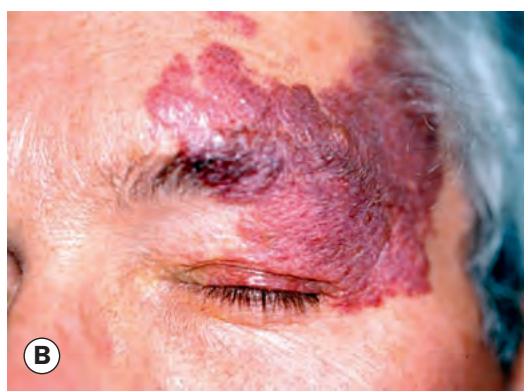
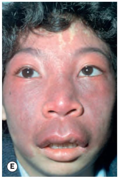
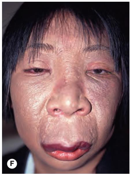

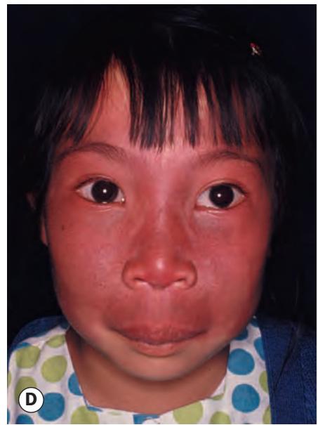
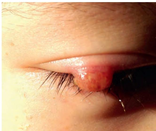

Fig. 1.14 Port-wine stain. (A) Histopathology shows widely dilated blood-filled spaces separated by fibrous septa; (B) and (C) *clinical appearance; (D–F) progression of port-wine stain over time, with associated underlying soft tissue hypertrophy* (Courtesy of L Horton – fig. A)
Fig. 1.15 Pyogenic granuloma

Fig. 1.16 Plexiform neurofibroma – characteristic S-shaped upper lid
(Courtesy of J Harry)