
Fig. 1.11 Syringomas
(Courtesy of A Pearson)
ophthalmologists. It affects children and young adults and is more common in females. Clinically it appears as a mobile purplish dermal nodule that may have a hard consistency due to calcification (Fig. 1.12A). Histopathology shows irregular epithelial islands exhibiting viable basophilic cells at the periphery and degenerate ‘shadow’ cells more centrally (Fig. 1.12B). Calcification is frequently present and there is often a foreign body giant cell reaction. Treatment involves excision. Malignant change is rare. Other, less common, hair follicle proliferations include trichofolliculoma, trichoepithelioma and trichilemmoma.
MISCELLANEOUS BENIGN TUMOURS
Capillary haemangioma
Capillary haemangioma (strawberry naevus) is one of the most common tumours of infancy; it is three times as common in boys as girls. It presents shortly after birth as a unilateral, raised bright red lesion (Fig. 1.13A), usually in the upper lid; a deeper lesion appears purplish (Fig. 1.13B and see also Fig. 3.31). Ptosis is frequent. The lesion blanches on pressure and may swell on crying. There may be orbital extension (see Ch. 3). Occasionally the lesion may involve the skin of the face and some patients have strawberry naevi on other parts of the body. Histopathology shows proliferation of varying-sized vascular channels in the dermis and subcutaneous tissue (Fig. 1.13C). It is important to be aware of an association between multiple cutaneous lesions and visceral haemangiomas, and to consider systemic assessment in appropriate cases. Treatment is described in Ch. 3.
Port-wine stain
Introduction
Port-wine stain (naevus flammeus) is a congenital malformation of vessels within the superficial dermis, consisting histopathologically of vascular spaces of varying calibre separated by thin fibrous septa (Fig. 1.14A). About 10% have associated ocular or CNS involvement, including Sturge–Weber (see below) and other defined syndromes.
Diagnosis
Port-wine stain manifests clinically as a sharply demarcated soft pink patch that does not blanch with pressure, most frequently located on the face. It is usually unilateral and tends to be aligned with the skin area supplied by one or more divisions of the trigeminal nerve (Figs 1.14B and C). Darkening to red or purple takes place with age, and there is commonly associated soft tissue hypertrophy (Figs 1.14D–F). Bleeding may occur from focal overlying lobulations (pyogenic granulomas – see below).
Treatment
Treatment with laser (e.g. pulsed-dye) is effective in decreasing skin discoloration; cosmetically superior results are usually achieved by early treatment. Topical preparations such as imiquimod and rapamycin, alone or with adjuvant laser, show promise. Soft tissue debulking is used in a small number of cases. Screening for glaucoma should begin in infancy. Systemic investigation is considered in some patients, particularly those with a lesion of the lumbar area.
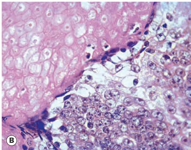
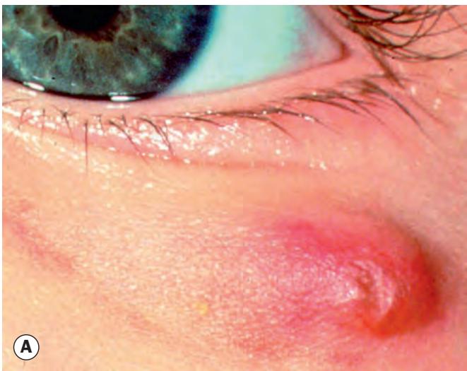
Fig. 1.12 Pilomatricoma. (A) Clinical appearance; (B) histopathology shows viable basophilic cells to the right and degenerate ‘shadow’ cells to the left
(Courtesy of J Krachmer, M Mannis and E Holland, from Cornea, Elsevier 2005 – fig. A; J Harry and G Misson, from Clinical Ophthalmic Pathology, Butterworth-Heinemann 2001 – fig. B)
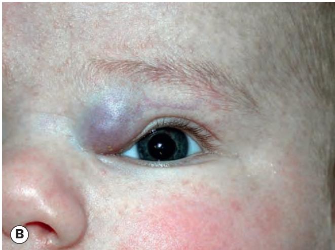
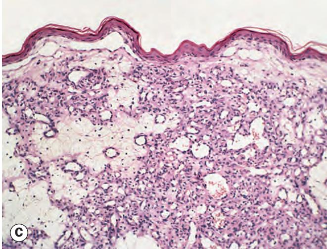
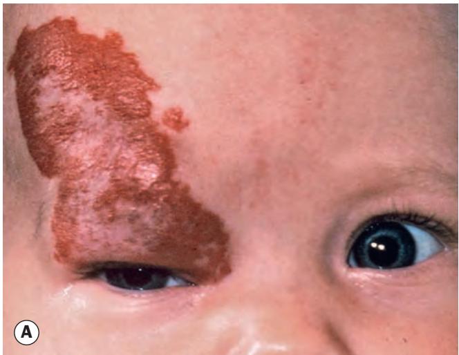
Fig. 1.13 Capillary haemangioma. (A) Medium-sized haemangioma; (B) mechanical ptosis due to a large lesion; (C) histopathology shows vascular channels of varying size within the dermis and subcutaneous tissue
(Courtesy of S Chen – fig. A; J Harry – fig. C)