Seborrhoeic keratosis
Seborrhoeic keratosis (basal cell papilloma) is an extremely common slowly growing lesion found on the face, trunk and extremities of elderly individuals as a discrete light- to dark-brown plaque with a friable, greasy, verrucous surface and a ‘stuck-on’ appearance (Fig. 1.6A). They are frequently numerous. The differential diagnosis includes pigmented basal cell carcinoma, naevus and melanoma. Histopathology shows expansion of the squamous epithelium of the epidermis by proliferating basal cells, sometimes with keratin-filled horns or cystic inclusions (Fig. 1.6B). Treatment involves shave biopsy (occasionally simple excision), electrodesiccation with curettage, laser ablation, cryotherapy with liquid nitrogen, and chemical peeling.
Actinic keratosis
Actinic (solar, senile) keratosis is a common slowly growing lesion that rarely develops on the eyelids. It typically affects elderly, fair-skinned individuals on areas of sun-damaged skin such as the forehead and backs of the hands, and appears as a hyperkeratotic plaque with distinct borders and a scaly surface that may become fissured (Fig. 1.7A). Occasionally the lesion is nodular or wart-like and may give rise to a cutaneous horn. Histopathology shows irregular dysplastic epidermis with hyperkeratosis, parakeratosis and cutaneous horn formation (Fig. 1.7B). It has potential, though low, for transformation into squamous cell carcinoma. Treatment involves biopsy followed by excision or cryotherapy.
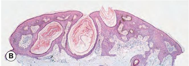

Fig. 1.6 Basal cell papilloma. (A) Typical ‘stuck-on’ appearance; (B) histopathology showing an elevated expansion of the epidermis with proliferation from basal cells – horn cysts and pseudohorn cysts are present
(Courtesy of A Pearson – fig. A; J Harry – fig. B)
BENIGN PIGMENTED LESIONS
Freckle
A freckle (ephelis, plural ephelides) is a small (generally 1–5 mm) brown macule due to increased melanin in the epidermal basal layer, typically in sun-exposed skin (Fig. 1.8); numbers vary with the level of sun exposure and can sometimes regress completely. Histopathology shows hyperpigmentation of the basal layer of the epidermis, with a normal melanocyte population.
Congenital melanocytic naevus
Congenital naevi are uncommon and histologically resemble their acquired counterparts (see below). They are usually small and of uniform colour. Rare variants include a ‘kissing’ or split naevus that involves the upper and lower eyelid (Fig. 1.9A) and may occasionally contain numerous hairs (Fig. 1.9B), and a very large lesion covering an extensive area of the body (“giant hairy naevus” – Fig. 1.9C). Large lesions have the potential for malignant transformation (up to 15%). Treatment, if necessary, involves complete surgical excision.
Acquired melanocytic naevus
Diagnosis
The clinical appearance and potential for malignant transformation of naevi are determined by their histological location within the skin.
Junctional naevus occurs in young individuals as a uniformly brown macule or plaque (Fig. 1.10A). The naevus cells are located at the junction of the epidermis and dermis and have a low potential for malignant transformation (Fig. 1.10B).
Compound naevus occurs in middle age as a raised papular lesion. The shade of pigment varies from light tan to dark
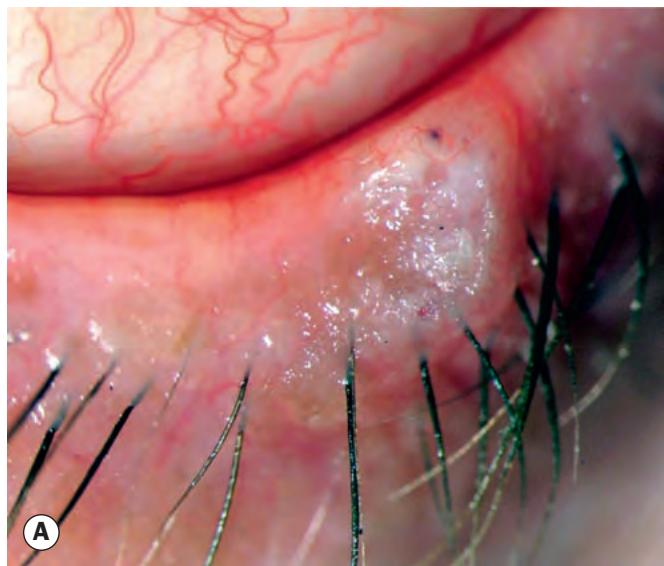
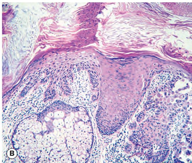
Fig. 1.7 Actinic keratosis. (A) Clinical appearance; (B) histopathology shows irregular dysplastic epidermis with hyperkeratosis, parakeratosis and cutaneous horn formation
(Courtesy of M Jager – fig. A; J Harry and G Misson, from Clinical Ophthalmic Pathology, Butterworth-Heinemann 2001 – fig. B)
Benign Adnexal Tumours
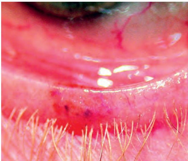
Fig. 1.8 Freckle (ephelis)
brown but tends to be relatively uniform throughout (Fig. 1.10C). The naevus cells extend from the epidermis into the dermis (Fig. 1.10D). It has a low malignant potential related to the junctional component.
Intradermal naevus, the most common, typically occurs in older patients. It is a papillomatous lesion, with little or no pigmentation (Fig. 1.10E). Histologically, naevus cells are confined to the dermis and have essentially no malignant potential (Fig. 1.10F).
Variants of naevi include balloon cell naevi, halo naevi, Spitz naevi (juvenile melanomas) and dysplastic naevi (atypical moles). Multiple dysplastic naevi constitute the dysplastic naevus syndrome (atypical mole syndrome – AMS). Individuals with AMS are at increased risk of developing conjunctival and uveal naevi and cutaneous, conjunctival and uveal melanomas.