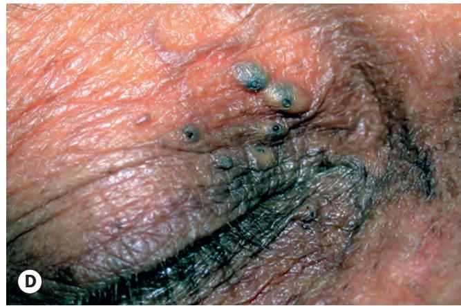
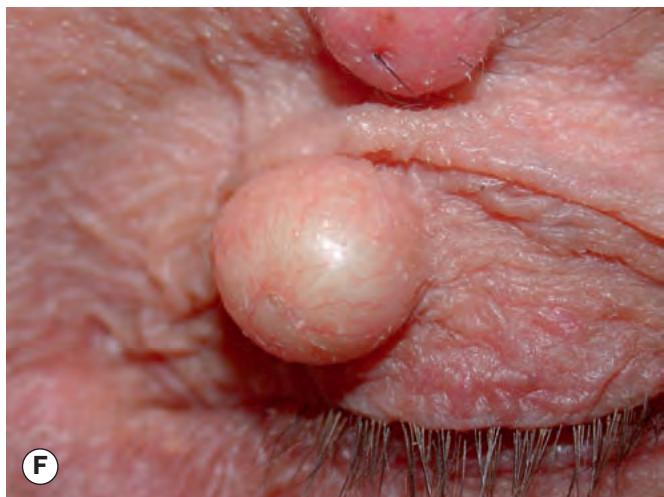
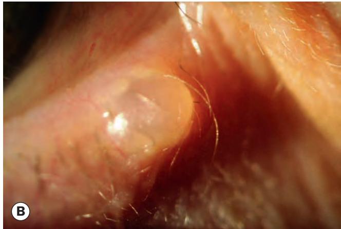
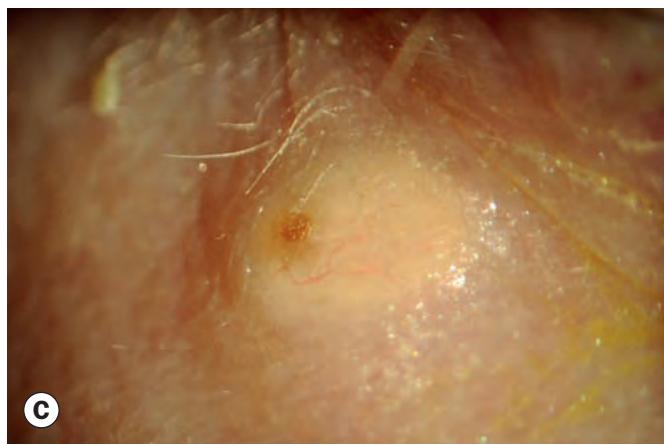
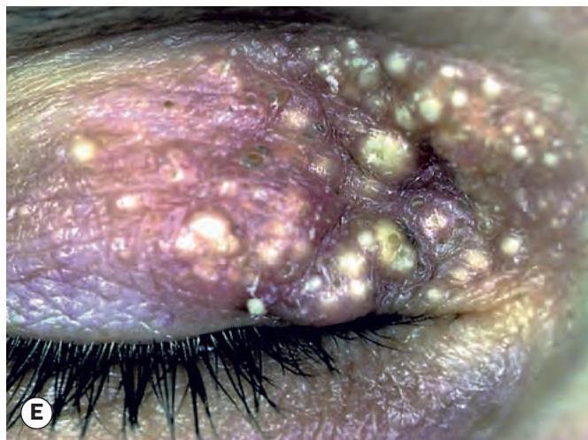
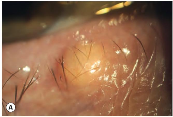
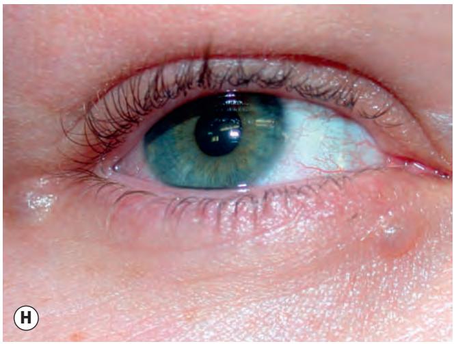
Fig. 1.3 Eyelid cysts. (A) Cyst of Zeis; (B) cyst of Moll; (C) sebaceous cyst; (D) comedones – blackheads; (E) milia; (F) epidermal inclusion cyst;
Xanthelasma
Introduction
Xanthelasma (plural – xanthelasmata) is a common, frequently bilateral condition typically affecting middle-aged and elderly individuals. It is a subtype of xanthoma. Hyperlipidaemia is found in about one-third of patients, in whom corneal arcus may also be present. In contrast to chalazion, fat in xanthelasmata is mainly intracellular, with lipid-laden histiocytes (foam cells) in the dermis (Fig. 1.4A).
Diagnosis
Xanthelasmata are yellowish subcutaneous plaques, usually in the medial aspects of the eyelids (Fig. 1.4B), commonly bilateral and are multiple (Fig. 1.4C).

Fig. 1.3, Continued (G) dermoid cyst; (H) eccrine hidrocystomas
(Courtesy of A Pearson – figs D, F and H) ablation and cryotherapy have advantages but may be more prone to scarring, including pigmentary changes.
Treatment
This is principally for cosmesis. Recurrence occurs in up to 50%, and is most common in patients with hypercholesterolaemia.
Simple excision is commonly performed where adequate excess skin is present.
Microdissection. Larger lesions can be raised in a flap, the fatty deposits dissected from overlying skin under a surgical microscope using microscissors, and the skin replaced.
Other methods. Good results can be obtained using chemical peeling with bi- or trichloroacetic acid. Laser