General considerations
Classification. Epidermal, adnexal or dermal.
Diagnosis. The clinical characteristics of benign lesions are a tendency to a lack of induration and ulceration, uniform colour, limited growth, regular outline and preservation of normal lid margin structures. Biopsy may be required if the appearance is suspicious.
○ Incisional biopsy involves removal of a portion of a lesion for histopathology.
○ Excision biopsy is performed on small tumours and fulfils both diagnostic and treatment objectives.
Treatment options include:
○ Excision of the entire lesion and a small surrounding portion of normal tissue.
☐ Marsupialization involves the removal of the top of a cyst allowing drainage of its contents and subsequent epithelialization.
○ Ablation with laser or cryotherapy.
NON-NEOPLASTIC LESIONS
Chalazion
Pathogenesis
A chalazion (meibomian cyst) is a sterile chronic granulomatous inflammatory lesion (lipogranuloma) of the meibomian, or sometimes Zeis, glands caused by retained sebaceous secretions. Histopathology shows a lipogranulomatous chronic inflammatory picture with extracellular fat deposits surrounded by lipid-laden epithelioid cells, multinucleated giant cells and lymphocytes (Fig. 1.2A). Blepharitis is commonly present; rosacea can be associated with multiple and recurrent chalazia. A recurrent chalazion should be biopsied to exclude malignancy.
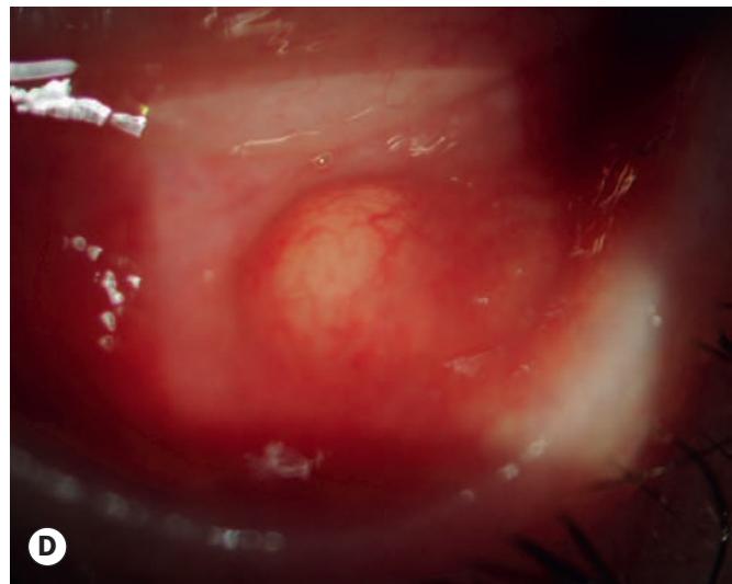
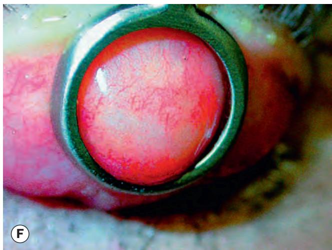

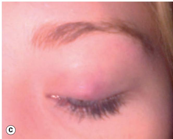

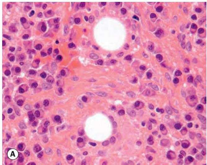
Fig. 1.2 Chalazion. (A) Histopathology shows a lipogranuloma; the large pale cells are epithelioid cells and the well-demarcated empty space contained fat dissolved out during processing; (B) uninflamed chalazion; (C) acutely inflamed lesion; (D) conjunctival granuloma; (E) marginal chalazion; (F) conjunctival view of chalazion clamp in place prior to incision and curettage
(Courtesy of J Harry and G Misson, from Clinical Ophthalmic Pathology, Butterworth-Heinemann 2001 – fig. A; J Nerad, K Carter and M Alford, from ‘Oculoplastic and Reconstructive Surgery’, in Rapid Diagnosis in Ophthalmology, Mosby 2008 – fig. F)