Dermis
The dermis is much thicker than the epidermis. It is composed of connective tissue and contains blood vessels, lymphatics and nerve fibres in addition to fibroblasts, macrophages and mast cells; upward dermal extensions (papillae) interdigitate with downward epidermal projections (rete ridges). In the eyelid the dermis lies on the orbicularis muscle. Adnexa lie deep in the dermis or within the tarsal plates.
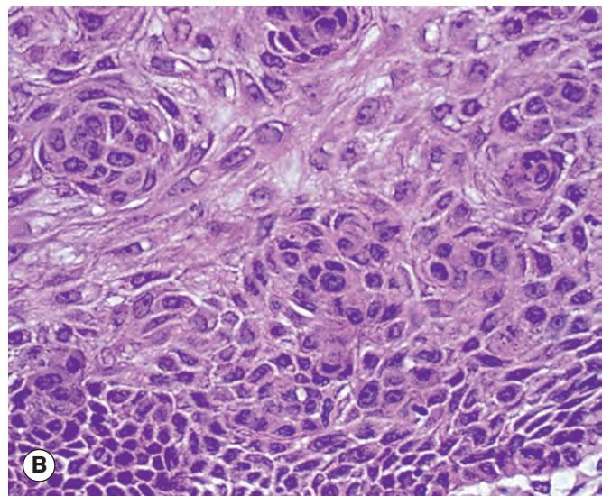


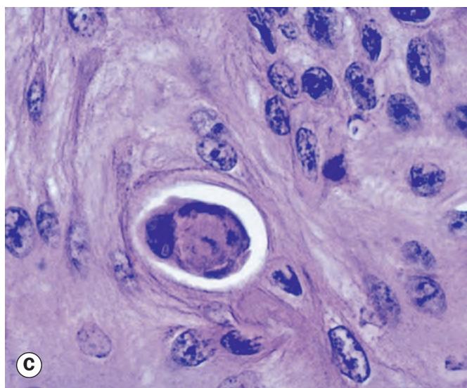
Fig. 1.1 Eyelid skin. (A) Normal skin is composed of keratinized stratified epithelium that covers the surface; pilosebaceous elements are conspicuous in the dermis and a few blood vessels and sweat glands are also seen; (B) dysplasia with loss of cell polarity; (C) dyskeratosis – a non-surface epithelial cell producing keratin; (D) parakeratosis – retention of cell nuclei into the surface keratin layer
(Courtesy of J Harry – fig. A; J Harry and G Misson, from Clinical Ophthalmic Pathology, Butterworth-Heinemann, 2001 – figs B–D)
Sebaceous glands are located in the caruncle and within eyebrow hairs. Tiny sebaceous glands are associated with the thin (vellus) hairs covering periocular skin.
Meibomian glands are modified sebaceous glands found in the tarsal plates. They empty through a single row of 20–30 orifices on each lid. A gland consists of a central duct with multiple acini, the cells of which synthesize lipids (meibum) that form the outer layer of the tear film.
Glands of Zeis are modified sebaceous glands associated with lash follicles.
Glands of Moll are modified apocrine sweat glands opening either into a lash follicle or directly onto the anterior lid margin between lashes; they are more numerous in the lower lid.
Eccrine sweat glands are distributed throughout eyelid skin and are not confined to the lid margin, in contrast to glands of Moll.
Pilosebaceous units comprise hair follicles and their sebaceous glands (see Fig. 1.1A).
Terminology
Clinical
Macule. Localized area of colour change without infiltration, depression or elevation, less than 1 cm in diameter.
Papule. A solid elevation less than 1 cm in diameter.
Vesicle. Circumscribed lesion containing serous fluid; less than 0.5 cm across.
Bulla. A large (more than 0.5 cm) serous fluid-filled lesion; plural – bullae.
Pustule. A pus-filled elevation less than 1 cm in diameter.
Crust. Solidified serous or purulent exudate.
Nodule. A palpable solid area measuring more than 1 cm.
Cyst. A nodule consisting of an epithelial-lined cavity filled with fluid or semi-solid material.
Plaque. A solid elevation of the skin, greater than 1 cm in diameter.
Scale. Readily detached fragments of shed keratin layer.
Papilloma. A benign neoplastic warty or tag-like projection of the skin or mucous membrane.
Ulcer. A circumscribed area of epithelial loss; in skin an ulcer extends through the epidermis into the dermis.
Histological
Tumour strictly refers only to a swelling, though is commonly used to denote a neoplasm.
Neoplasia. Abnormal tissue growth, either benign (localized, non-invasive and non-spreading) or malignant
(progressive growth with the potential for distant spread).
Atypia refers to an abnormal appearance of individual cells, e.g. abnormal mitotic figures.
Dysplasia is an alteration of the size, morphology and organization of cellular components of a tissue. There is disturbance of normally structured and recognized layers of tissue (e.g. loss of cell polarity – Fig. 1.1B).
Carcinoma in situ (intraepidermal carcinoma, Bowen disease) exhibits dysplastic changes throughout the thickness of the epidermis.
Hyperkeratosis. An increase in thickness of the keratin layer that appears clinically as scaling. Hyperkeratosis can be a feature of benign or malignant epithelial tumours.
Acanthosis. Thickening of the prickle cell layer.
Dyskeratosis is keratinization other than on the epithelial surface (Fig. 1.1C).
Parakeratosis is the retention of nuclei into the keratin layer (Fig. 1.1D).