Other Tests for Evaluating Suppression
Many other tests are available for testing suppression; these include both subjective and objective tests. Commonly used subjective tests include the AO (American Optical) vectographic chart, the near Mallett unit, Bagolini striated lenses, and cheiroscopic tracings. The 4 base-out test is an objective method of assessing suppression. We suggest the use of the Worth four-dot test because of its availability, low cost, ease of administration, and accuracy in detecting suppression. A complete discussion of instrumentation and specific clinical procedures is available in other texts (44,45).
EVALUATION OF STEREOPSIS
Randot Stereotest
1. Purpose The Randot Stereotest is a subjective test designed to evaluate the presence and degree of stereopsis at near using both global and contour (local) stereopsis targets.
Important issues
(a) Global versus contour targets Two techniques are commonly used for the assessment of stereopsis. The first, called contour or local stereopsis, uses two similar targets that are laterally displaced. The Titmus stereofly, Wirt rings, and animals (Fig. 1.3) are examples of this type of target. A shortcoming of this type of stereopsis target is that patients with no stereopsis may be able to guess the “correct” answer using monocular cues. Cooper and Warshowsky (46) found that the correct response for the first four Wirt rings could be determined by looking for monocular displacement of one of the circles. Clinically this may be significant when a clinician is examining a child who is trying to give the “right answer” to please the examiner. Of course, with a patient giving accurate responses and not trying to fool the examiner, this test works well.
The second type of stereopsis technique, called global targets, eliminates this problem. Global targets contain random dot stereopsis targets and have no monocular cues. As a result, the guessing that can occur with contour stereopsis is not a problem with global stereopsis.
Another important distinction between contour and global targets is their value in detecting the presence of a constant strabismus. Cooper and Feldman (47) investigated the use of stereopsis tests to detect strabismus and found that with a random dot stereogram of 660 seconds of arc disparity, no constant strabismic could pass the test. Thus, even a gross random dot stereopsis target is effective at detecting the presence of a constant strabismus. With contour stereopsis targets, a constant strabismic can occasionally appreciate up to 70 seconds of arc stereopsis (43). Random dot targets can be used to rule out the presence of a constant strabismus, whereas contour stereopsis targets can be used to determine whether peripheral stereopsis is present. Peripheral stereopsis is considered to be any value greater than 60 seconds of arc. Both types of stereopsis targets, therefore, have value and it is best to use both in the clinical evaluation of stereopsis. This can be accomplished using one test, such as the Randot Stereotest (Fig. 1.4A), or by using two tests, such as the Synthetic Opticsa circle, square, and E targets (Fig. 1.4B) or butterfly (Fig. 1.4C) and the Titmus Stereotest or the Synthetic Optics animals and circles.1 (b) Polaroid versus anaglyphic targets The traditional evaluation of stereopsis includes measurement by stimulation of retinal disparity using polarized targets and polarized glasses. Targets not requiring the use of any glasses have also been developed, and stereopsis measurements with these targets have been shown to correlate well with those requiring polarized glasses (48). Another format for measuring stereopsis involves the use of red/green cancellation to induce exclusive, disparate images to the right and left eyes. Thus, red/green testing is often referred to as anaglyphic testing. These targets have been developed so that other target properties, such as disparity, shape, and size, are similar to those found in the polarized equivalents. Yamada et al. (49) found that the red/green method yields results comparable to the polarized equivalent, especially for testing the presence of random dot or global stereopsis in patients. The red/green version of the random dot butterfly/butterfly stereopsis test and the random dot letter “E”/RDE tests (Synthetic Opticsa) offer a cost-effective alternative for clinicians when attempting to rule out the presence of constant strabismus. To measure the level of contour stereopsis, their data suggest lower agreement between red/green and polarized methods. Therefore, if the objective is to quantify improvement of contour stereopsis after treatment, polarized versions of stereopsis tests may be more useful.

Figure 1.3 A: Titmus stereofly. B: Child reaching for “fly” that appears to be floating in front of the page.
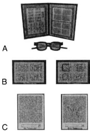
Figure 1.4 Examples of random dot stereopsis. A: Randot Stereotest. B: The Synthetic Optics circle, square. C: The Synthetic Optics butterfly target.
(c) Near versus distance stereopsis testing Most clinicians routinely evaluate stereopsis using targets designed for use at 33 cm or 40 cm. In most cases this is sufficient because if stereopsis is present at 40 cm it should be present at distance as well. The exception would be a case in which there is an intermittent or constant strabismus at distance. In such cases the clinician should consider testing stereopsis at distance as well. Currently there are both noncomputer- and computer-based tests for distance stereopsis. An example of a computer-based assessment is the M&S $^{\circledR}$ Technologies Vision Testing System that has a distance stereopsis test which uses LCD Shutter Glasses. It is likely that other computerized systems will also incorporate distance stereopsis testing. A new noncomputer-based distance stereo assessment is the Distance Randot Test (Stereo Optical). Wang et al. (50) reported that the Distance Randot scores from normal subjects have low variability and high test- retest reliability. They concluded that the Distance Randot Stereotest is a sensitive measurement of binocular sensory status that may be useful in monitoring progression of strabismus and/or recovery following strabismus surgery.
1Throughout the book, see end-of-chapter lists for sources of equipment identified by superscript letters in the text.
3. Expected values A patient with normal binocular function should be able to achieve 20 seconds of stereopsis with the contour stimuli and appreciate stereopsis with the gross random dot targets.