The maximum plus refraction technique breaks down when acuity is very unequal (e.g., amblyopia). In these instances, where often no refractive technique works well, use retinoscopy to determine balance after attempting to achieve maximum plus on the “good” eye (make the retinoscopic reflexes appear equal for the two eyes).
Assessment of Nonstrabismic Binocular Vision Disorders
GENERAL CONSIDERATIONS
The evaluation of binocular vision involves several distinct steps (Table 1.1). The first phase of testing is the measurement of the magnitude and direction of the phoria at a distance and near, along with the accommodative convergence to accommodation (AC/A) ratio. Conventional procedures to accomplish this include tests such as cover testing, the von Graefe phoria test, and the modified Thorington test. Fixation disparity testing represents a more recent method of assessing binocular vision and provides additional information that should be considered in the evaluation of binocular vision status. The primary advantage of fixation disparity testing is that it is performed under binocular or associated conditions, in contrast to other tests that are performed under dissociated conditions.
The second step is the assessment of positive and negative fusional vergence using both direct and indirect measures. Direct measures refer to tests such as smooth and step vergence testing, whose primary objective is to assess fusional vergence. Indirect measures refer to tests such as the negative relative accommodation (NRA), positive relative accommodation (PRA), fused cross-cylinder, binocular accommodative facility (BAF), and monocular estimation method (MEM) retinoscopy that are generally thought of as tests of accommodative function. Because these procedures are performed under binocular conditions, however, they indirectly evaluate binocular function as well. The results of such testing, therefore, can be used to confirm or deny a particular clinical hypothesis of a binocular vision disorder. Chapter 2 describes the analysis of these indirect measures in detail.
The traditional evaluation of fusional vergence involves only measurement of smooth vergence ranges or vergence amplitude using a Risley prism in the phoropter. In recent years, additional ways of evaluating fusional vergence have been suggested. One method is step vergence testing, which is done outside the phoropter, using a prism bar (1,2). Another addition to the traditional approach to assessing fusional vergence is vergence facility testing (3–9). This test is also performed outside the phoropter, using a specially designed vergence facility prism (Fig. 1.1). The patient’s ability to make large rapid changes in fusional vergence is assessed with this procedure over a specific period of time.
An important distinction among different methods of evaluating fusional vergence is the assessment of vergence amplitude versus vergence facility. Smooth and step vergence testing are designed to assess the patient’s vergence amplitude, whereas vergence facility testing measures vergence dynamics. Grisham (6) found a relationship between vergence dynamics and symptoms in subjects he studied. His research indicated that vergence latency and vergence velocity are of diagnostic importance in a binocular evaluation. It is possible for a patient to have normal fusional vergence amplitudes and still have a problem in the area of facility or vergence dynamics. Using only the traditional smooth vergence evaluation approach would fail to detect such a problem. Gall et al. (7) found that the use of 3Δ base-in/12 Δ base-out for vergence facility testing can differentiate symptomatic from nonsymptomatic patients.
TABLE 1.1 Important Steps in the Evaluation of Binocular Vision
| Measurement of the phorias | Cover test |
| AC/A and CA/C ratios | von Graefe phoria Modified Thorington Fixation disparity |
| Assessment of positive and negative fusional vergence | |
| Direct measures | Smooth vergence testing Step vergence testing Vergence facility testing |
| Indirect measures | Negative relative accommodation Positive relative accommodation Fused cross-cylinder Binocular accommodative facility Monocular estimation method retinoscopy |
| Convergence amplitude Sensory status | Near point of convergence Worth four-dot Stereopsis testing |
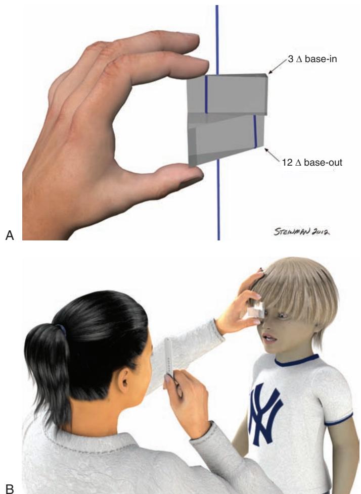
Figure 1.1 A: Vergence facility prism (3 $ \Delta $ base-in/12 $ \Delta $ base-out). B: Vergence facility prism clinical procedure.
Another consideration in testing fusional vergence amplitude or facility is the issue of performance over time (3). The underlying question is whether the patient is able to compensate for a given amount of prism over an extended period of time. Traditionally, fusional vergence amplitude is measured just once. Research suggests that this may not be sufficient (6,7). Rather, these tests should be repeated several times, and testing that probes facility and ability to respond over time should be incorporated into the evaluation.
The third area that should be evaluated is convergence amplitude. Generally referred to as the near point of convergence (NPC), this test is particularly important in the diagnosis of one of the most common binocular vision disorders—convergence insufficiency. Important issues include the type of target or targets to be used and the issue of performance over time (10,11).
The last aspect of the binocular evaluation is sensory status. Suppression and stereopsis are the primary areas to evaluate. Information about sensory status can also be obtained from many of the other tests discussed above. On several of these tests, suppression can be monitored. A specific test that can be used to assess suppression is the Worth four-dot test. As a general rule, clinical measures of stereopsis are either not affected or only minimally affected in nonstrabismic binocular vision disorders. Intermittent mild suppression, however, is a common finding.
A complete assessment of binocular vision should include all four of the components just described. A suggested minimum database would include the NPC, the cover test at distance and near, step vergence ranges at distance and near, and stereopsis testing. If a patient presents with symptoms and the minimum database does not yield conclusive information, additional testing using indirect measures of binocular function, along with facility testing and fixation disparity assessment, should be utilized.