The corneal topography demonstrates regular with-the-rule astigmatism, which appears as a vertically oriented symmetric bowtie. According to the SimK values, there is 2.13 D of astigmatism in the 80 degree meridian OD and 4.50 D of astigmatism in the 80 degree meridian OS.
Corneal astigmatism tends to be with-the-rule in children and typically decreases with age. Adults tend to have against-the-rule astigmatism that increases with age. Approximately 50% of the population has 0.75 D or more of astigmatism. The total astigmatism in the eye is the sum of that in the cornea (anterior and posterior surfaces [posterior contributes approximately 0.4 D against-the-rule]), the lens, and rarely the retina.
Keratorefractive: incisions (astigmatic keratotomy, corneal/limbal relaxing incisions) with a blade or laser, ablation (PRK, LASIK), and lenticule extraction (SMILE).
Lenticular: toric IOL (phakic IOL or pseudophakic IOL).
7
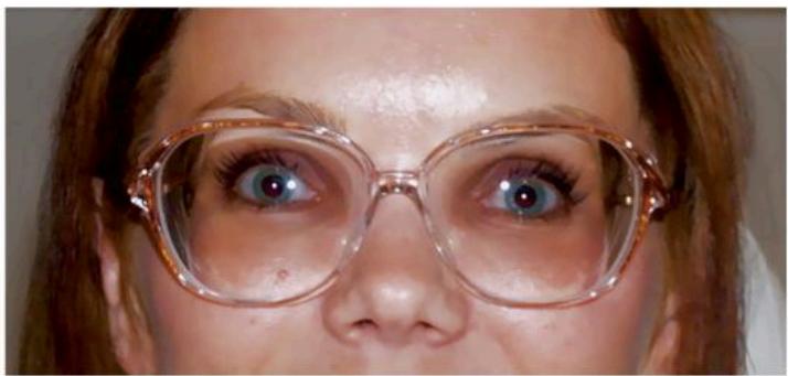
A 38-year-old myopic woman complains that her bulky, heavy glasses are a nuisance and she would like to investigate other options.
What are the alternatives and what other information would you like to know?
Additional information: she has worn glasses for 30 years, the last change was 7 years ago, she is not interested in contact lenses, has no other eye problems, and has not noticed a recent change in vision. Her current glasses are - 7.25 + 1.00 × 100OD and - 8.00 + 2.50 × 20OS, her vision with these glasses is 20/20OU, her refraction yields the same prescription and vision. There is no evident anterior or posterior segment abnormality on slit-lamp exam and ophthalmoscopy. She is interested in refractive surgery.
What surgical options are available and what tests would you perform?
Additional information: corneal pachymetry is thinnest centrally and measures 505~μm OD and 510~μm OS, angles are open/grade 4 OU, anterior chamber depth (ACD) is 3.5~mm OD and 3.6~mm OS, and endothelial cell count is normal OU. Her corneal topography shows:
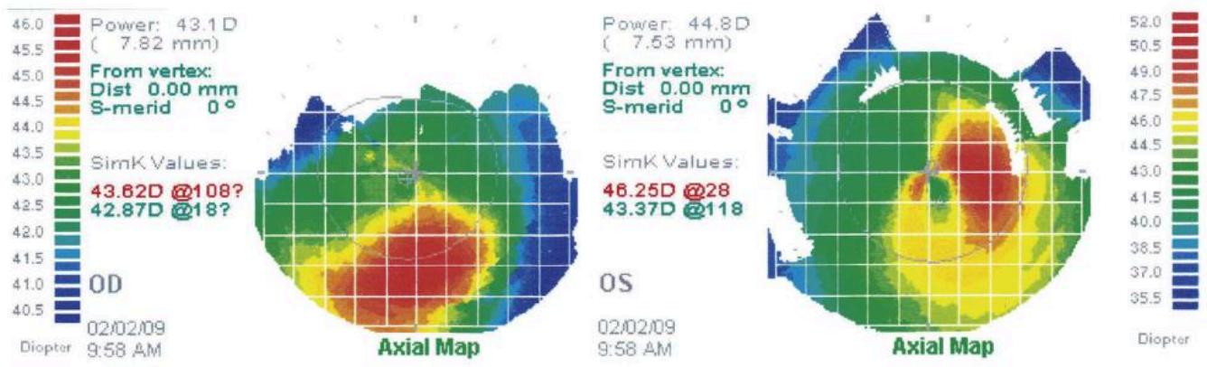
How do you interpret the topography maps, and how do the new data affect her surgical options?
What are the complications of phakic IOL implantation?
Depending on her refractive error, her options include: new glasses with high-index lenses, contact lenses, and refractive surgery. It would be important to know more about her ocular history and exam. Specifically, when did she start wearing glasses, when was her last prescription change, has she tried contact lenses, and if so what type, for how long, and what did she think about them? Does she have any other past ocular history? Has her vision changed recently? How do the glasses interfere with her lifestyle, daily activities, and hobbies? Has she thought about refractive surgery and, if so, what does she hope to accomplish? With regard to the eye exam, what is her current prescription, visual acuity with correction and manifest refraction? Are there any abnormalities on exam?
Possible surgical options are laser vision correction, phakic IOL, and RLE. However, the disadvantages of RLE are the loss of accommodation and greater risk of vision-threatening complications due to more invasive surgery. To assist in deciding which option is most appropriate, it is necessary to obtain corneal topography, pachymetry, gonioscopy, ACD, and endothelial cell evaluation.
The computerized videokeratoscopy reveals forme fruste keratoconus OD and mild pellucid marginal degeneration OS, and her corneas are thin, so she is not a candidate for laser vision correction. Her surgical options are lens procedures: phakic IOL or RLE. Since she has a stable refraction and excellent vision with glasses at age 38 years old, it is unlikely that her corneas will change significantly. If she desires phakic IOL surgery, a toric design should be considered to correct her astigmatism OS since corneal astigmatic procedures (corneal-relaxing incisions and laser vision correction) should not be performed. However, a small amount of residual ametropia could possibly be corrected with PRK, particularly after corneal collagen cross-linking. Similarly, if she prefers RLE, then toric presbyopia-correcting lenses or toric monofocal IOLs set for monovision should be considered, otherwise she will require glasses for the presbyopia or astigmatism. Multifocal IOLs are usually not a good choice in eyes with this type of abnormal corneal topography because the irregular corneal astigmatism may cause some degree of natural multifocality, increases the unpredictability of IOL calculations, and precludes postoperative keratorefractive surgical enhancement to correct residual ametropia. However, if the irregular astigmatism is mild, is regular/symmetric centrally, the visual acuity is not reduced, and the patient is happy with a multifocal SCL trial, then a presbyopia-correcting IOL can be considered.
The main risks are corneal endothelial cell loss, glaucoma, iritis, and cataract. The risk of these complications varies depending on the style of IOL:
Angle-supported: highest risk of corneal endothelial damage; may also develop glaucoma.
Iris-supported: highest risk of iris damage, iritis, and IOL dislocation.
Posterior chamber: highest risk of cataract and angle-closure glaucoma.
Furthermore, there is a small risk of lens decentration/dislocation, infection, cystoid macular edema, retinal detachment, and disruption of the anterior lens capsule.
8
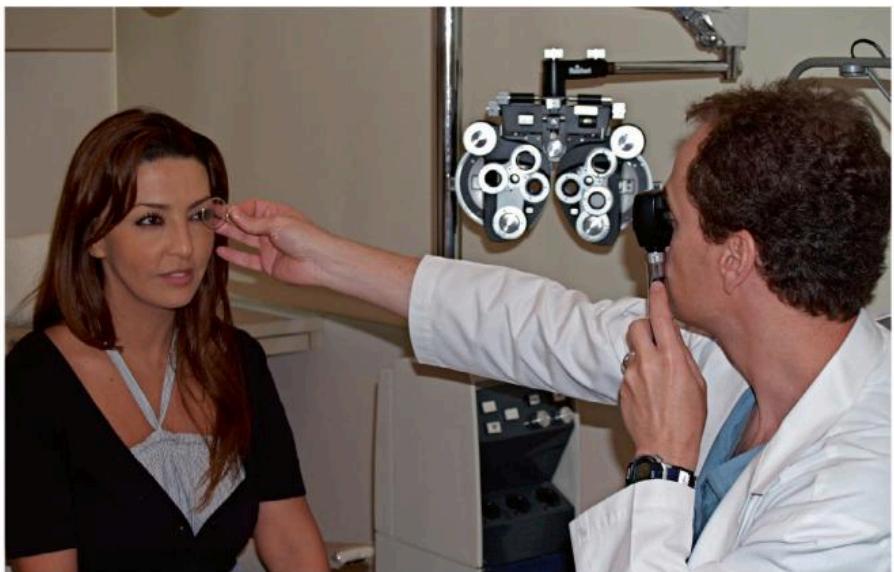
A 34-year-old woman broke her glasses and wants a new prescription. She does not have any old records or glasses to measure.
Describe the technique of streak retinoscopy.
What methods can be used for an irregular cornea?
Retinoscopy is a method of objectively measuring the refractive state of an eye. With a phoropter or free lenses, the examiner moves the light streak across the pupil and changes the correcting lenses until the reflex movement is neutralized (pupil appears uniformly illuminated). This occurs at the far point. If the far point is between the examiner and patient (myopic), the reflex moves in the direction opposite to the retinoscope sweep (“against” motion). If the far point is behind the examiner (hyperopia), the reflex has “with” motion. The final refraction is then determined by adjusting for the working distance (add reciprocal of working distance to the final finding).
For a scissoring reflex, which occurs in keratoconus, observe the central area of the reflex. If the reflex is poor or irregular, then alternate methods should be used, such as a contact lens overrefraction or stenopeic slit refraction.