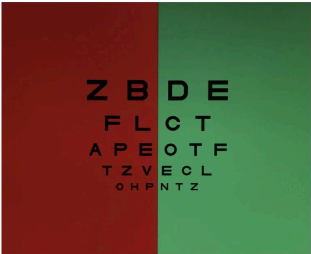
What is the name of the test depicted in the photo?
How does this test work and how is it performed?
The duochrome test.
The duochrome test uses the principle of chromatic aberration to monocularly refine the spherical endpoint and check the accuracy and balance of the refraction. Light of shorter wavelengths is refracted more than light of longer wavelengths (green is bent more than red and is therefore focused anterior to red). The green and red filters create a 0.5 D difference. The patient must therefore have 20/30 or better visual acuity in order to distinguish the difference. If the patient sees the letters on the green side of the chart clearer, then the focal point is behind the retina (the eye is overminused) and plus sphere is added to the prescription. If the patient sees the red side clearer, then the focal point is in front of the retina (the eye is overplussed) and minus sphere is added to the prescription. This can be remembered with the mnemonic RAM-GAP: Red Add Minus - Green Add Plus. The technique is to start with the red side clearer and add minus sphere in 0.25 D steps until the letters on the red and green sides are of equal clarity (ie, the red and green light are the same distance (0.25 D) behind and in front of the retina, respectively), then the focal point is on the retina and the prescription is balanced. The duochrome test works in colorblind patients because it uses chromatic aberration, not color discrimination.
20
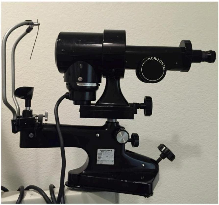
What device is shown and how does it work?
What are the limitations of this device?
What is the astigmatic effect from the posterior corneal surface?
A keratometer. This device measures the curvature of the anterior corneal surface based on the power of a reflecting surface. It does this by measuring the size of an image reflected from 2 paracentral points and utilizes doubling prisms to stabilize the image enabling more accurate focusing. The anterior corneal curvature is then calculated using the convex mirror formula, and the corneal power is estimated empirically based on Snell’s law of refraction with simplified optics. The keratometer adjusts the index of refraction (1.3375 versus 1.376) to account for the posterior corneal power and also to allow 45 D to equal 7.5 mm radius of curvature (K [diopters] = 337.5/r).
Limitations of the keratometer include: it only measures a small region of the cornea (2 points at the 3–4 mm zone), it does not provide information about the cornea central or peripheral to these points, it measures different regions for corneas of different powers, it assumes the cornea is spherocylindrical and symmetric with a major and minor axis separated by 90 degrees, it ignores spherical aberration, it is susceptible to focusing and misalignment errors, and it cannot accurately measure irregular corneas.
The posterior corneal surface contributes approximately 0.4 D of against-the-rule astigmatism to the total corneal power. This is important when determining the amount of corneal astigmatic correction during cataract surgery and RLE.
21
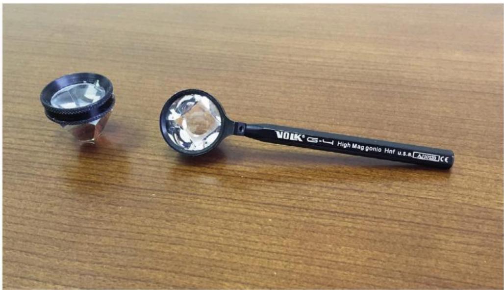
Name the various types of gonioscopy lenses and explain how they differ.
Define the critical angle.
What is total internal reflection and how is this relevant to the eye exam?
There are a variety of gonioscopy lenses: Koeppe-style lenses (ie, Koeppe, Barkan, Wurst, Swan-Jacob, and Richardson) for direct gonioscopy (direct and upright view of the iridocorneal angle structures with a binocular microscope, light source, and supine patient, usually in the operating room), and the Goldmann-style lenses and the 4- or 6-mirror lenses (ie, Zeiss, Posner, Sussman) for indirect gonioscopy (indirect and inverted view of the opposite angle with the slit lamp biomicroscope and patient sitting upright).
The Goldmann lenses typically contain three mirrors, but there are lenses with only one mirror. The lens rests on the sclera, vaults over the cornea and therefore requires a viscous coupling solution (ie, Goniosol), and cannot be used for indentation gonioscopy. In the three-mirrors lenses, each mirror has a different angulation to view different parts of the eye: the angle ( ∼ 60 degrees), peripheral retina ( ∼ 65 degrees), and mid-peripheral retina ( ∼ 75 degrees). The center of the lens can be used to visualize the posterior pole. Because only one of the mirrors can be used for angle viewing, the lens must be rotated 360 degrees to examine the entire angle.
The 4- and 6-mirror lenses are composed of symmetric mirrors with the same angulation ( ∼ 60–63 degrees) so that the angle structures can be viewed with minimal or no rotation of the lens. These lenses rest on the cornea without the need for a coupling solution. This enables the examiner to perform indentation gonioscopy to differentiate between appositional and synechial angle closure. Appositional angle closure opens with indentation of the central cornea, which pushes aqueous peripherally, and synechial angle closure does not open with indentation.
The critical angle is the angle at which incident light is bent 90 degrees away from the normal (when going from a medium of higher to lower index of refraction, n) and after which all light is reflected. The critical angle differs for different interfaces and depends upon the indices of refraction of the material on either side of the interface. The critical angle of the cornea (air/cornea interface) is 46.5 degrees.
Total internal reflection is an optical phenomenon that occurs when the angle of incidence exceeds the critical angle. This causes light to be reflected back into the material with higher index of refraction. Angle structures cannot be directly visualized owing to total internal reflection at the air/cornea interface. Therefore, a gonioscopy lens is required to view the angle structures.