Measure refraction and keratometry.
Choose base curve steeper than þat K (usually +0.50 D steeper to form a tear lens; tear lens prevents apical touch).
Convert refraction to minus cylinder form and zero vertex distance; disregard the cylinder (minus cylinder is formed by the tears).
Power of contact lens is sphere from refraction adjusted for tear lens (subtract +0.50); “SAM-FAP.”
To evaluate the fit, assess the fluorescein pattern, lens movement, and centration.
The power of SCL is based on the spherical equivalent manifest refraction corrected for vertex distance. The base curve is based on average keratometry measurements. To evaluate the fit, assess the movement of the lens. Poor movement means the lens is too tight (too steep) and excessive movement indicates that the lens is too flat.
The lens is too tight, so to loosen it either increase the radius of curvature or decrease the diameter.
16
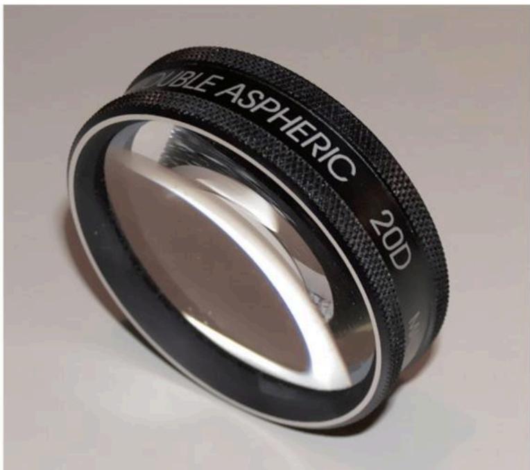
Condensing lenses are used with an indirect ophthalmoscope to examine the retina.
What are the advantages of the indirect ophthalmoscope?
What are the different types of magnification?
What is the retinal magnification with a 20 D lens?
Larger field of view and stereopsis compared with the direct ophthalmoscope. Field of view is 25 degrees (versus 7 degrees) and magnification is approximately 2–3× depending on the lens (versus 15×).
Transverse, axial, and angular magnification.
Transverse (linear or lateral): magnification of image size (away from the optical axis). Equal to the ratio of image height to object height ( ML = I/O).
Axial: magnification of depth (along the optical axis). Equal to the square of the transverse magnification ( MAX = ML2).
Angular: magnification of angle subtended by an image with respect to an object. Used when the object or image size cannot be measured. ( MA=D/4, standardized to 25 cm [1/4 m], the near point of the average eye.)
MA=Deye/Dlens=60/20=3×.
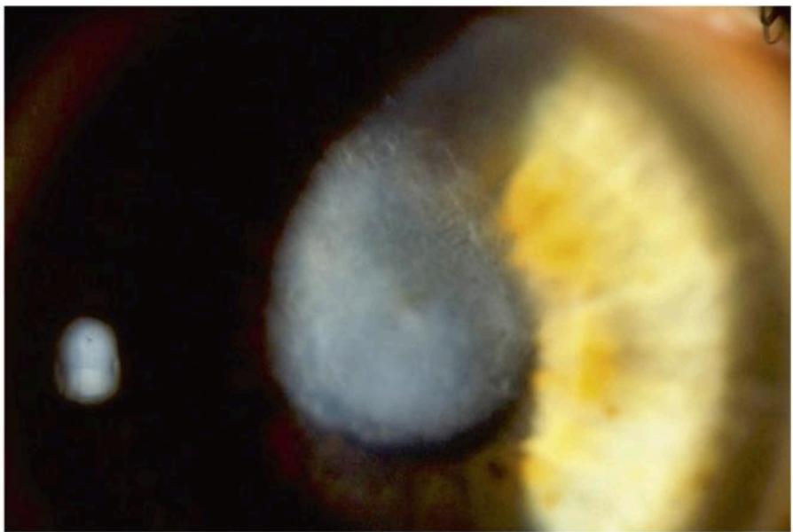
A 34-year-old man with a corneal scar and BSCVA of 20/100 in his right eye wants to know if his vision can be improved.
How would you evaluate him?
If this patient has better visual potential, what are the surgical options?
A number of tests can be performed to determine the visual potential including pinhole acuity, RGP contact lens overrefraction, and PAM test. VER could also be performed.
Depending on the location, size, and depth of the scar, the options are PTK, rotational corneal autograft, penetrating keratoplasty, and ALK, superficial SALK, or deep DALK.
18
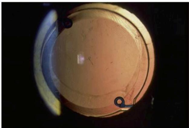
A 68-year-old man sees you for a second opinion. He complains of blurred vision since having cataract surgery 2 months ago. On exam, his uncorrected vision is 20/100 at distance and J1 at near in the operated eye. He is unhappy because he expected good distance vision without glasses and would like to wear glasses to read.
What is the problem?
What history and findings would be helpful?
What are the possible etiologies?
Additional information: old records reveal a history of myopia ( 7.50 D), RGP contact lens wear until age 46 when he had LASIK surgery OU. Corneal topography shows:
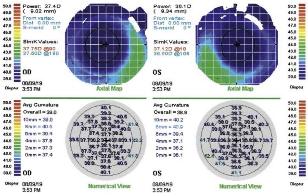
What is the most likely cause of his refractive surprise?
What are the surgical treatment options?
Myopic refractive outcome.
The patient should be asked about previous use of glasses/contact lenses, magnitude of prescription, ocular surgery (ie, refractive and retinal procedures) or injuries. Old records are valuable if available. Helpful exam findings include incisions or scarring from previous surgery or trauma, integrity of the capsular bag, IOL position and stability, status of the macula and peripheral retina, and signs of myopic fundus.
A refractive surprise after cataract surgery occurs because the IOL is the wrong power or is in the wrong position. Wrong IOL power is due to an error in AL measurement, error in keratometry measurement, use of an incorrect IOL calculation formula, or insertion of the wrong IOL. Wrong IOL position is due to sulcus placement without reducing the IOL power, and anterior displacement of an IOL in the bag from a large capsulorhexis, capsular distension/capsular block syndrome, capsular contraction, or upside-down placement of an angulated IOL.
Corneal topography shows a well-centered myopic LASIK ablation with very flat keratometry readings, which is a significant factor in IOL calculation error. There is often a wide range of IOL formula results, which makes choosing the correct IOL power difficult. Intraoperative wavefront aberrometry is helpful in eyes with previous corneal refractive surgery.
This patient can be treated with IOL exchange, piggyback IOL, and laser vision correction. For capsular block syndrome, a Nd:YAG laser anterior capsulotomy should be performed to release the trapped fluid and allow the IOL to return to its normal position.