Incisional Techniques
Incisions perpendicular to the corneal surface alter its shape, depending on the direction, depth, location, length, and number of incisions (see Chapter 3). All incisions cause a local flattening of the cornea. Radial incisions lead to flattening in both the meridian of the incision and the one 90° away. Tangential (arcuate or linear) incisions lead to flattening in the meridian of the incision and steepening in the meridian 90° away that may be equal to or less than the magnitude of the decrease in the primary meridian (Fig 1-20); this phenomenon is known as coupling (see Chapter 3, Fig 3-3). Reducing the optical zone of radial incisions increases their effect; similarly, placing tangential incisions closer to the visual axis increases the effect, as does increasing the length of a tangential incision, up to 3 clock-hours.
For optimum effect, an incision should be 85%–90% deep to retain an intact posterior lamella and maximum anterior bowing of the other lamellae. Nomograms for the number of incisions and optical zone size can be calculated using finite element analysis, but sur gical nomograms are typically generated empirically (eg, see Chapter 3, Table 3-1). The important variables for radial and astigmatic surgery include patient age and the number, depth, and length of incisions. The same incision has greater effect in older patients than it does in younger patients. IOP and preoperative corneal curvature are not significant predictors of effect.
Technological developments have led to the use of femtosecond lasers for making arcuate incisions. Advanced imaging capabilities allow creation of femtosecond laser–assisted astig matic or arcuate keratotomy (AK) with precise arc length, depth, and location (Video 1-1). Incorporation of iris registration technology allows cyclotorsion compensation and facilitates accurate placement of AK incisions on the intended meridian. Femtosecond laser–assisted AKs have been documented to yield good outcomes with low risks of wound dehiscence or corneal perforation.

VIDEO 1-1 Femtosecond laser astigmatic keratotomy post–penetrating keratoplasty.
Courtesy of Karolinne Maia Rocha, MD, PhD.


Figure 1-20 Schematic diagrams of incisions used in astigmatic keratotomy. Flattening is induced in the axis of the incisions (at $ 90^{\circ} $ in this case), and steepening is induced $ 90^{\circ} $ away from the incisions (at $ 180^{\circ} $ in this case). (Illustrations by Cyndie C. H. Wooley.)
Arcuate
Tangential
Limbal relaxing incisions
Chan TC, Cheng GP, Wang Z, Tham CC, Woo VC, Jhanji V. Vector analysis of corneal astigmatism after combined femtosecond-assisted phacoemulsification and arcuate keratotomy. Am J Ophthalmol. 2015;160(2):250–255.e252.
Chang JSM. Femtosecond laser-assisted astigmatic keratotomy: a review. Eye Vis (Lond). 2018;5:6.
Rowsey JJ. Ten caveats in keratorefractive surgery. Ophthalmology. 1983;90(2):148–155.
Visco DM, Bedi R, Packer M. Femtosecond laser-assisted arcuate keratotomy at the time of cataract surgery for the management of preexisting astigmatism [published correction appears in J Cataract Refract Surg. 2020;46(4):658]. J Cataract Refract Surg. 2019;45(12):1762–1769.
Tissue Addition or Subtraction Techniques
With the exception of laser ablation techniques (discussed in the section Laser Biophys ics), lamellar procedures that alter corneal shape through tissue addition or subtraction are primarily of historical interest. Keratomileusis for myopia was originated by Barraquer as “carving” of the anterior surface of the cornea. It is defined as a method to modify the spherical or meridional surface of a healthy cornea by tissue subtraction. Epikeratoplasty (sometimes called epikeratophakia) adds a precision-lathed lenticule of donor tissue to the corneal surface to induce hyperopic or myopic changes. Keratophakia requires the addi tion of a tissue lenticule or synthetic inlay intrastromally.
However, there is renewed interest in tissue subtraction as a way to treat refractive error without excimer laser ablation. These current procedures—called refractive lenticule extraction (ReLEx), femtosecond lenticule extraction (FLEx), and small-incision lenticule *extraction (SMILE)—*use a femtosecond laser to excise an intrastromal lenticule and thus alter the corneal curvature.
Alloplastic Material Addition Techniques
The shape of the cornea can be altered by adding alloplastic material such as hydrogel on the surface or in the corneal stroma to modify the anterior shape or refractive index of the cornea. For example, the 2 arc segments of an intrastromal corneal ring can be placed in 2 pockets of the stroma to directly reshape the surface contour according to the profile and location of the individual segments (Fig 1-21; see also BCSC Section 8, External Disease and Cornea).
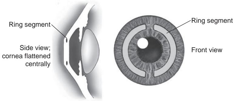
Figure 1-21 Schematic illustrations showing placement of intrastromal corneal ring segments. (Illustrations by Jeanne Koelling.)
A different type of inlay, which alters optical function by means of a small-aperture effect, was approved for treatment of presbyopia in 2015. Although other styles of corneal inlays and onlays have been investigated for presbyopia, studies were halted; in one case, an approved product was withdrawn from the market because of high rates of corneal haze. For further discussion, see Chapter 10.