Evaluation Methods
Diagnostic techniques that directly evaluate biomechanics before refractive surgery, ide ally to help differentiate between healthy and abnormal corneas, are available and in de velopment. Devices currently available rely on bidirectional applanation tonometry or corneal deformation measurement with Scheimpflug imaging.
With noncontact bidirectional applanation, a focused air jet is used to indent the cor nea and record 2 pressure measurements: the pressure at which the cornea moves inward, reaching the first applanation, and the pressure at which the cornea recovers from a slight concavity as the air pump decreases pressure at an inverse rate (the second applanation). The pressure at the second applanation is lower than that at the first; this difference is called corneal hysteresis (CH), which represents the viscous (time-dependent) dissipation of energy in the cornea (Fig 1-19, left). Low CH has been associated with keratoconus (Fig 1-19, right) and identified as an independent risk factor for progressive visual field loss in glaucoma. The mean of these 2 pressures is the Goldmann-correlated IOP (IOPg). The corneal-compensated IOP (IOPcc) is a pressure measurement that takes into account the biomechanical properties and is independent of the central corneal thickness. The corneal resistance factor (CRF) is an indicator of the overall resistance to the air puff that is weighted more toward the instantaneous (elastic) properties of the cornea.
The corneal deformation method uses an ultra-high-speed Scheimpflug camera to mon itor the deformation of the cornea from a noncontact tonometer air-puff indentation.
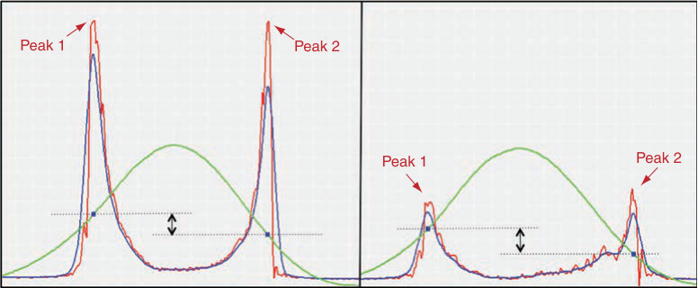
Figure 1-19 Noncontact bidirectional applanation. The device delivers a collimated stream of air onto the corneal apex (green curve); the optical signal (red curve) represents the dynamic motion of the cornea during the rapid in/out deformation. The filtered applanation signals are plotted in blue (blue line). The difference between the inward and outward pressure values defines corneal hysteresis. (Left) Repeatable high-amplitude peaks with minimal signal noise are seen in normal eyes. (Right) Low-amplitude peaks and signal noise are observed in kerato conus patients. (Courtesy of Karolinne Maia Rocha, MD, PhD.)
This method provides detailed information about the biomechanical characteristics of the cornea by determining velocity, length, and time lapse during different phases of applanation.
Abahussin M, Hayes S, Knox Cartwright NE, et al. 3D collagen orientation study of the human cornea using x-ray diffraction and femtosecond laser technology. Invest Ophthalmol Vis Sci. 2009;50(11):5159–5164.
Blackburn BJ, Jenkins MW, Rollins AM, Dupps WJ. A review of structural and biomechanical changes in the cornea in aging, disease, and photochemical crosslinking. Front Bioeng Biotechnol. 2019;7:66.
Damgaard IB, Reffat M, Hjortdal J. Review of corneal biomechanical properties following LASIK and SMILE for myopia and myopic astigmatism. Open Ophthalmol J. 2018;12:164–174.
Dupps WJ Jr, Wilson SE. Biomechanics and wound healing in the cornea. Exp Eye Res. 2006;83(4):709–720.
Garcia-Porta N, Fernandes P, Queiros A, Salgado-Borges J, Parafita-Mato M, González- Méijome JM. Corneal biomechanical properties in different ocular conditions and new measurement techniques. ISRN Ophthalmol. 2014;2014:724546.
Glass DH, Roberts CJ, Litsky AS, Weber PA. A viscoelastic biomechanical model of the cornea describing the effect of viscosity and elasticity on hysteresis. Invest Ophthalmol Vis Sci. 2008;49(9):3919–3926.
Changes in Corneal Biomechanics After Refractive Procedures
Corneal biomechanical properties are of major importance in keratorefractive surgeries. The various procedures differ in the biomechanical response they produce, depending on the biomechanical load of the ablated or dissected cornea. In LASIK, a corneal flap is created with a microkeratome or femtosecond laser, followed by thinning of the stromal bed with excimer laser ablation. In photorefractive keratectomy (PRK), the laser is ap plied directly to the anterior corneal stroma without creating a flap. In small-incision lenticule extraction (SMILE), a femtosecond laser is used to create a 3-dimensional len ticule, which is then extracted through a small corneal incision ranging from 2 to 5 mm. Because no flap is created and the most anterior stromal lamellae remain intact (except in the region of the small incision), this procedure may provide better biomechanical stability. Some studies have demonstrated better CH and CRF measurements following SMILE compared with LASIK or laser subepithelial keratomileusis (LASEK), possibly because the stiffer anterior stroma is preserved. However, ectasia has been reported after SMILE, as well.
Chen M, Yu M, Dai J. Comparison of biomechanical effects of small incision lenticule extraction and laser-assisted subepithelial keratomileusis. Acta Ophthalmol. 2016;94(7): e586–e591.
Dou R, Wang Y, Xu L, Wu D, Wu W, Li X. Comparison of corneal biomechanical characteristics after surface ablation refractive surgery and novel lamellar refractive surgery. Cornea. 2015;34(11):1441–1446.
Ganesh S, Brar S, Arra RR. Refractive lenticule extraction small incision lenticule extraction: a new refractive surgery paradigm. Indian J Ophthalmol. 2018;66(1):10–19.
Sachdev G, Sachdev MS, Sachdev R, Gupta H. Unilateral corneal ectasia following small- incision lenticule extraction. J Cataract Refract Surg. 2015;41(9):2014–2018.
Wang D, Liu M, Chen Y, et al. Differences in the corneal biomechanical changes after SMILE and LASIK. J Refract Surg. 2014;30(10):702–707.
Wu D, Wang Y, Zhang L, Wei S, Tang X. Corneal biomechanical effects: small-incision lenticule extraction versus femtosecond laser-assisted laser in situ keratomileusis. J Cataract Refract Surg. 2014;40(6):954–962.
Corneal Effects of Keratorefractive Surgery
All keratorefractive procedures induce refractive changes by altering corneal curva ture; however, the method by which the alteration is accomplished varies by the pro cedure and the refractive error. Treatment of myopia requires a flattening, or decrease, in central corneal curvature, whereas treatment of hyperopia requires a steepening, or increase, in central corneal curvature. Keratorefractive surgery can be performed using a variety of techniques, including incisional, tissue addition or subtraction, alloplastic material addition, and laser ablation (see the section Laser Biophysics for discussion of laser ablation).
Overall patient satisfaction after the surgery depends largely on the successful correc tion of refractive error and creation of a corneal shape that maximizes visual quality. As discussed previously, the normal prolate corneal shape produces an aspheric optical sys tem, while oblate corneas increase sphere aberration and visual symptoms such as glare, halos, and reduced contrast sensitivity. (See the section “Effect of excimer laser ablation on higher-order aberrations.”)