Corneal Biomechanics
Characteristics of the Stroma
The corneal stroma is the dominant structural component of the cornea and consists of colla gen fibrils arranged in approximately 200 lamellar sheets. This network of collagen is largely responsible for the mechanical strength of the cornea. Lamellae are oriented parallel to the corneal surface at alternating angles to the adjacent lamellae, and the predominant fiber orientations in the posterior two-thirds of the stroma are vertical and horizontal. The fibrils demonstrate more branching and interweaving in the anterior third of the stroma, which confers greater biomechanical strength to the anterior stroma. In keratoconus, branching fibers are sparse in the area of the cone, which may cause local weakness. Structural differ ences between the anterior and posterior stroma are relevant in assessing the relative impact of procedures such as LASIK, PRK, and SMILE.
The type and distribution of glycosaminoglycans differ between the anterior and posterior stroma, and the more hydrophilic proteins in the posterior stroma influence
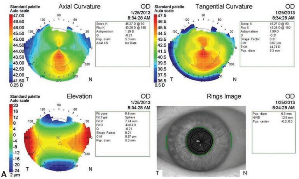
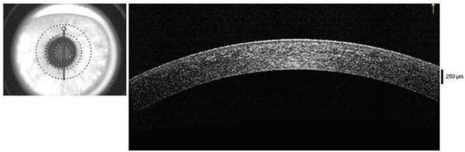
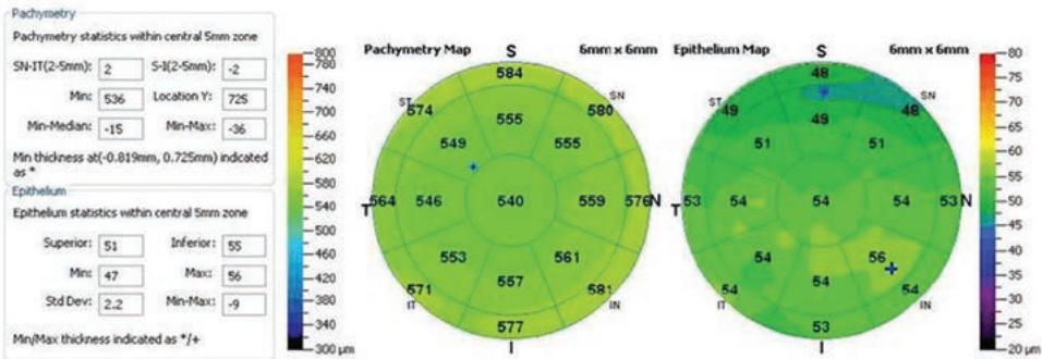
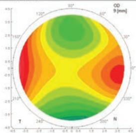
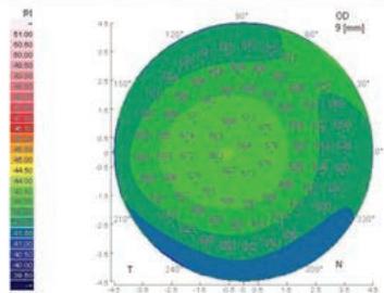
Figure 1-15 Different options for corneal imaging. All images are of the same patient taken at the same visit. A, Placido disk–based corneal curvature map showing axial and tangential curvature maps as well as the elevation map and the Placido mires. This mapping technol ogy analyzes only the surface characteristics of the cornea. B, Optical coherence tomography image. Note that the corneal thickness profile (of the stroma as well as the epithelium) is well demonstrated, but the overall surface curvature is not.
(Continued)
Axial curvature
[01.2275 R000+8.0 pmq]
Anterior elevation
R00+6.0 [mm] BFS radius=7.68 [mm]
Corneal pachymetry
RON=9.8 [puro]
Posterior elevation

R048+7.8 [mm] EFS radius=6.61 [mm]
C