For routine refractive screening, most surgeons use the axial curvature mode rather than the instantaneous mode for topographic output.
Corneal topography and astigmatism
A topographic image of a normal cornea without astigmatism demonstrates a relatively uni form color pattern centrally with a natural flattening in the periphery (Fig 1-13A). Regular astigmatism is uniform steepening along a single corneal meridian that can be fully corrected with a cylindrical lens. Topographic imaging of regular astigmatism demonstrates a sym metric “bow-tie” pattern along a single meridian with a straight axis on both sides of center (Fig 1-13B). The bow-tie pattern on topographic maps is an artifact of Placido-based imag ing; that is, because the Placido image cannot detect curvature at the central measurement point, the corneal meridional steepening seems to disappear centrally and become enhanced farther from center.
Irregular astigmatism is nonuniform corneal steepening from a variety of causes that can not be corrected by cylindrical lenses. Irregular astigmatism decreases corrected distance vi sual acuity (CDVA) and may also reduce contrast sensitivity and increase visual aberrations, depending on the magnitude of irregularity. Rigid gas-permeable contact lenses can correct visual acuity reductions resulting from corneal irregular astigmatism by bridging the irregu lar corneal surface and the contact lens with the tear film. For more information on irregular astigmatism, see BCSC Section 3, Clinical Optics.
Corneal topography is very helpful in evaluating eyes with irregular astigmatism. Topographic changes include nonorthogonal steep and flat meridians (ie, not 90° apart; Fig 1-14). Asymmetry between the superior and inferior or nasal and temporal halves of the cornea may also be revealed by corneal topography, although these patterns are not necessarily indicative of corneal pathology. In contrast, wavefront analysis can demonstrate higher-order aberrations such as coma, trefoil, quadrafoil, or secondary astigmatism. The ability to differentiate regular from irregular astigmatism has clinical significance in keratorefractive surgery. Traditional excimer laser ablation can correct spherocylindrical errors but does not effectively treat irregular astigmatism. Topography-guided ablation may be useful in treating irregular astigmatism not caused by early corneal ectatic disorders.
Ghoreishi M, Naderi Beni A, Naderi Beni Z. Visual outcomes of topography-guided excimer laser surgery for treatment of patients with irregular astigmatism. Lasers Med Sci. 2014;29(1):105–111.
Lin DT, Holland S, Tan JC, Moloney G. Clinical results of topography-based customized ablations in highly aberrated eyes and keratoconus/ectasia with cross-linking. J Refract Surg. 2012;28(11 Suppl):S841–S848.
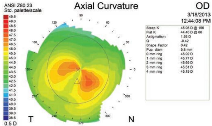
Figure 1-14 A curvature map showing nonorthogonal axes, which may indicate pathology that would contraindicate refractive surgery. (Courtesy of Gregg J. Berdy, MD.)
Limitations of corneal topography
In addition to the limitations of specific algorithms and the variations in terminology among manufacturers, the accuracy of corneal topography may be affected by other po tential problems:
tear film irregularities
misalignment (misaligned corneal topography may give a false impression of cor neal apex decentration suggestive of keratoconus)
instability (test-to-test variation)
insensitivity to focus errors
limited area of coverage (central and limbal)
decreased accuracy of corneal power simulation measurements (SIM K) after re fractive surgical procedures
decreased accuracy of posterior surface elevation values (with scanning-slit tech nology) in the presence of corneal opacities or, often, after refractive surgery
Roberts C. Corneal topography: a review of terms and concepts. J Cataract Refract Surg. 1996;22(5):624–629.
Corneal Tomography
Whereas surface corneal curvature (power) is best expressed by Placido-based topogra phy, overall corneal shape, including spatial thickness profiles, is best expressed by tomog raphy. Various imaging systems are available that take multiple slit images and reconstruct them into a corneal shape profile, including anterior and posterior corneal elevation data (Fig 1-15). Among the methods used are scanning-slit technology, Scheimpflug-based imaging systems, and anterior segment optical coherence tomography (OCT) that can provide epithelial mapping for refractive surgery. To represent shape directly, color maps may be used to display a z-height from an arbitrary plane such as the iris plane; however, in order to be clinically useful, corneal surface maps are plotted to show differences from best-fit spheres or other objects that closely mimic the normal corneal shape (Fig 1-16). In general, each device calculates the best-fit sphere individually for each map. For this reason, elevation maps cannot be compared precisely because they often have different referenced best-fit sphere characteristics.
Elevation-based tomography is especially helpful in refractive surgery for depicting the anterior and posterior surface shapes of the cornea and lens. With such information, alterations to the shape of the ocular structures, especially postoperative changes, can be determined with greater accuracy.
Indications for Corneal Imaging in Refractive Surgery
Corneal topography is an essential part of the preoperative evaluation of refractive surgery candidates. About two-thirds of patients with normal corneas have a symmetric astigma tism pattern that is round, oval, or bow-tie shaped (see Figure 1-13). Asymmetric patterns include asymmetric bow-tie patterns, inferior steepening, superior steepening, skewed radial axes, or other nonspecific irregularities.
Corneal topography detects irregular astigmatism, which may result from abnormal tear film, contact lens warpage, keratoconus and other corneal ectatic disorders, corneal surgery, trauma, scarring, and postinflammatory or degenerative conditions. Repeated topographic examinations may be helpful when the underlying etiology is in question, especially in cases of suspicious steepening patterns in patients who wear contact lenses or who have an abnormal tear film. It is often helpful to have contact lens users discon tinue their lens wear for an extended period before preoperative planning; this allows the corneal map and refraction to stabilize. Patients with keratoconus or other ectatic disor ders are not routinely considered for ablative keratorefractive surgery because the abnor mal cornea may exhibit an unpredictable response or progressive ectasia. Forme fruste, or subclinical, keratoconus typically is considered a contraindication to ablative refractive surgery. Studies are under way to determine the suitability of some keratorefractive pro cedures in combination with corneal crosslinking as alternative therapeutic modalities for these patients (see also BCSC Section 8, External Disease and Cornea).
Corneal topography and tomography can also be used to demonstrate the effects of keratorefractive procedures. Preoperative and postoperative maps may be compared to determine the refractive effect achieved (difference map; Fig 1-17). Corneal mapping can also help explain unexpected results, including undercorrection and overcorrection; in duced astigmatism; and induced aberrations from small optical zones, decentered abla tions, or central islands (Fig 1-18).
Ambrósio R Jr, Alonso RS, Luz A, Coca Velarde LG. Corneal-thickness spatial profile and corneal-volume distribution: tomographic indices to detect keratoconus. J Cataract Refract Surg. 2006;32(11):1851–1859.
De Paiva CS, Harris LD, Pflugfelder SC. Keratoconus-like topographic changes in kerato- conjunctivitis sicca. Cornea. 2003;22(1):22–24.
Rabinowitz YS, Yang H, Brickman Y, et al. Videokeratography database of normal human corneas. Br J Ophthalmol. 1996;80(7):610–616.