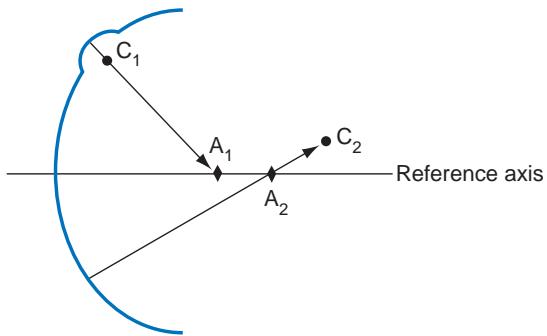
Figure 1-10 Schematic representation of the difference between axial distance (axial curvature) and radius of curvature for 2 points on a curved surface. Points $ \mathrm{C}_{1} $ and $ \mathrm{C}_{2} $ represent the centers of curvature of their respective surface points. Points $ \mathrm{A}_{1} $ and $ \mathrm{A}_{2} $ represent the endpoints of the axial distances for the given axis. Steeper, localized areas of curvature are underestimated, while flatter areas are overestimated. (Adapted from Roberts C. Corneal topography: a review of terms and concepts. J Cataract Refract Surg. 1996;22(5):624-629.)
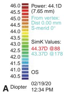
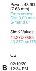
Figure 1-11 Examples of Placido-based curva ture maps. A, Axial (sagittal); B, instantaneous (tangential). (Courtesy of Karolinne Maia Rocha, MD, PhD.)

Instantaneous power and curvature
A second method of describing the corneal curvature on Placido disk–based topography is the instantaneous radius of curvature (also called meridional or tangential power). The in stantaneous radius of curvature is determined by taking a perpendicular path through the point in question from a plane that intersects the point and the visual axis, while allowing the radius to be the length necessary to correspond to a sphere with the same curvature at that point. The curvature, which is expressed in diopters, is estimated by the difference between the corneal index of refraction and 1.000, divided by this tangentially determined radius. A tangential map typically shows better sensitivity to peripheral changes, with less smoothing of the curvature than seen on an axial map (see Figures 1-11B and 1-12B). In these maps, diopters are relative units of curvature and are not the equivalent of diopters of corneal power. The potential benefit of this method’s greater sensitivity is balanced by its tendency to document excessive detail (“noise”), which may not be clinically relevant.
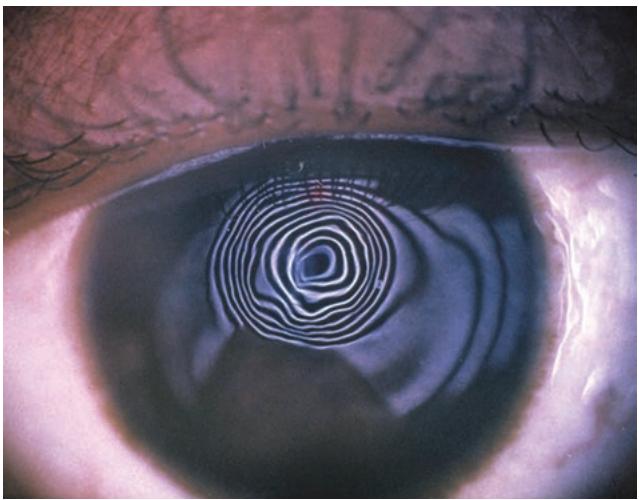
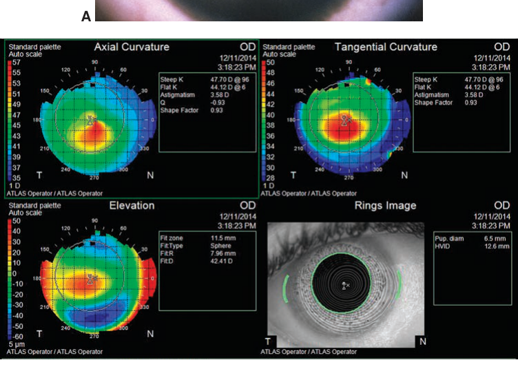
Figure 1-12 Corneal topography in keratoconus. A, Placido imaging showing distorted corneal mires. B, Axial, tangential, and elevation topography maps of the same cornea. (Courtesy of M. Bowes Hamill, MD.)
A
B
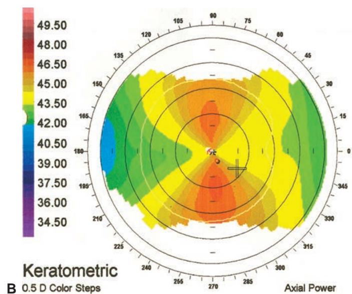

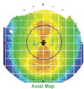
Figure 1-13 Common corneal topographic patterns. A, Prolate (normal); B, symmetric bow tie (regular astigmatism). (Courtesy of J. Bradley Randleman, MD.)