Методы оценки
Evaluation Methods
Существуют и разрабатываются диагностические методы, которые позволяют напрямую оценивать биомеханику перед рефракционной хирургией, что в идеале помогает дифференцировать здоровые и аномальные роговицы. Доступные в настоящее время устройства основаны на двунаправленной аппланационной тонометрии или измерении деформации роговицы с помощью визуализации по методу Шеймпфлюга.
Diagnostic techniques that directly evaluate biomechanics before refractive surgery, ide ally to help differentiate between healthy and abnormal corneas, are available and in de velopment. Devices currently available rely on bidirectional applanation tonometry or corneal deformation measurement with Scheimpflug imaging.
При использовании бесконтактной двунаправленной аппланационной тонометрии сфокусированная струя воздуха используется для вдавливания роговицы и регистрации 2 измерений давления: давления, при котором роговица смещается внутрь, достигая первой аппланации, и давления, при котором роговица восстанавливается после небольшой вогнутости по мере того, как воздушный насос снижает давление с обратной скоростью (вторая аппланация). Давление при второй аппланации ниже, чем при первой; эта разница называется роговичным гистерезисом (CH), который представляет собой вязкую (зависящую от времени) диссипацию энергии в роговице (Рис 1-19, слева). Низкий CH ассоциирован с кератоконусом (Рис 1-19, справа) и идентифицирован как независимый фактор риска прогрессирующей потери поля зрения при глаукоме. Среднее значение этих 2 давлений — это внутриглазное давление, коррелированное с методом Гольдмана (IOPg). Корнеально-компенсированное ВГД (IOPcc) — это измерение давления, которое учитывает биомеханические свойства и не зависит от центральной толщины роговицы. Коэффициент роговичного сопротивления (CRF) является показателем общего сопротивления воздушному импульсу, который в большей степени ориентирован на мгновенные (эластические) свойства роговицы.
With noncontact bidirectional applanation, a focused air jet is used to indent the cor nea and record 2 pressure measurements: the pressure at which the cornea moves inward, reaching the first applanation, and the pressure at which the cornea recovers from a slight concavity as the air pump decreases pressure at an inverse rate (the second applanation). The pressure at the second applanation is lower than that at the first; this difference is called corneal hysteresis (CH), which represents the viscous (time-dependent) dissipation of energy in the cornea (Fig 1-19, left). Low CH has been associated with keratoconus (Fig 1-19, right) and identified as an independent risk factor for progressive visual field loss in glaucoma. The mean of these 2 pressures is the Goldmann-correlated IOP (IOPg). The corneal-compensated IOP (IOPcc) is a pressure measurement that takes into account the biomechanical properties and is independent of the central corneal thickness. The corneal resistance factor (CRF) is an indicator of the overall resistance to the air puff that is weighted more toward the instantaneous (elastic) properties of the cornea.
Метод деформации роговицы использует ультравысокоскоростную камеру Шеймпфлюга для мониторинга деформации роговицы при воздействии воздушного импульса бесконтактного тонометра.
The corneal deformation method uses an ultra-high-speed Scheimpflug camera to mon itor the deformation of the cornea from a noncontact tonometer air-puff indentation.
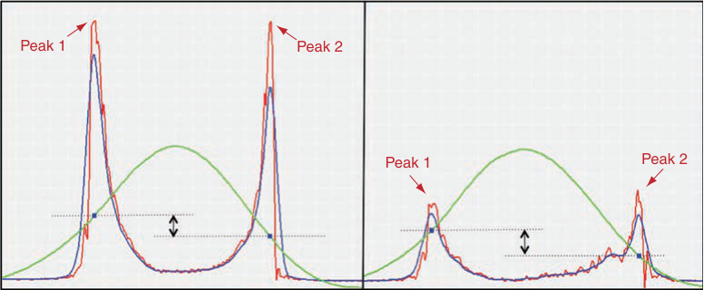
Рисунок 1-19 Бесконтактная двунаправленная аппланация. Устройство подает коллимированный поток воздуха на верхушку роговицы (зеленая кривая); оптический сигнал (красная кривая) представляет динамическое движение роговицы во время быстрого вдавливания/выталкивания. Отфильтрованные сигналы аппланации нанесены синим цветом (синяя линия). Разница между значениями давления при вдавливании и выталкивании определяет гистерезис роговицы. (Слева) В норме наблюдаются повторяющиеся высокоамплитудные пики с минимальным шумом сигнала. (Справа) У пациентов с кератоконусом наблюдаются низкоамплитудные пики и шум сигнала. (Предоставлено Karolinne Maia Rocha, MD, PhD.)
Figure 1-19 Noncontact bidirectional applanation. The device delivers a collimated stream of air onto the corneal apex (green curve); the optical signal (red curve) represents the dynamic motion of the cornea during the rapid in/out deformation. The filtered applanation signals are plotted in blue (blue line). The difference between the inward and outward pressure values defines corneal hysteresis. (Left) Repeatable high-amplitude peaks with minimal signal noise are seen in normal eyes. (Right) Low-amplitude peaks and signal noise are observed in kerato conus patients. (Courtesy of Karolinne Maia Rocha, MD, PhD.)
Этот метод предоставляет подробную информацию о биомеханических характеристиках роговицы путем определения скорости, длины и временного интервала во время различных фаз аппланации.
This method provides detailed information about the biomechanical characteristics of the cornea by determining velocity, length, and time lapse during different phases of applanation.
Abahussin M, Hayes S, Knox Cartwright NE, et al. 3D collagen orientation study of the human cornea using x-ray diffraction and femtosecond laser technology. Invest Ophthalmol Vis Sci. 2009;50(11):5159–5164.
Abahussin M, Hayes S, Knox Cartwright NE, et al. 3D collagen orientation study of the human cornea using x-ray diffraction and femtosecond laser technology. Invest Ophthalmol Vis Sci. 2009;50(11):5159–5164.
Blackburn BJ, Jenkins MW, Rollins AM, Dupps WJ. A review of structural and biomechanical changes in the cornea in aging, disease, and photochemical crosslinking. Front Bioeng Biotechnol. 2019;7:66.
Blackburn BJ, Jenkins MW, Rollins AM, Dupps WJ. A review of structural and biomechanical changes in the cornea in aging, disease, and photochemical crosslinking. Front Bioeng Biotechnol. 2019;7:66.
Damgaard IB, Reffat M, Hjortdal J. Review of corneal biomechanical properties following LASIK and SMILE for myopia and myopic astigmatism. Open Ophthalmol J. 2018;12:164–174.
Damgaard IB, Reffat M, Hjortdal J. Review of corneal biomechanical properties following LASIK and SMILE for myopia and myopic astigmatism. Open Ophthalmol J. 2018;12:164–174.
Dupps WJ Jr, Wilson SE. Биомеханика и заживление ран роговицы. Exp Eye Res. 2006;83(4):709–720.
Dupps WJ Jr, Wilson SE. Biomechanics and wound healing in the cornea. Exp Eye Res. 2006;83(4):709–720.
Garcia-Porta N, Fernandes P, Queiros A, Salgado-Borges J, Parafita-Mato M, González- Méijome JM. Биомеханические свойства роговицы при различных состояниях глаз и новые методы измерения. ISRN Ophthalmol. 2014;2014:724546.
Garcia-Porta N, Fernandes P, Queiros A, Salgado-Borges J, Parafita-Mato M, González- Méijome JM. Corneal biomechanical properties in different ocular conditions and new measurement techniques. ISRN Ophthalmol. 2014;2014:724546.
Glass DH, Roberts CJ, Litsky AS, Weber PA. A viscoelastic biomechanical model of the cornea describing the effect of viscosity and elasticity on hysteresis. Invest Ophthalmol Vis Sci. 2008;49(9):3919–3926.
Glass DH, Roberts CJ, Litsky AS, Weber PA. A viscoelastic biomechanical model of the cornea describing the effect of viscosity and elasticity on hysteresis. Invest Ophthalmol Vis Sci. 2008;49(9):3919–3926.
Изменения биомеханики роговицы после рефракционных процедур
Changes in Corneal Biomechanics After Refractive Procedures
Биомеханические свойства роговицы имеют первостепенное значение в кераторефракционной хирургии. Различные процедуры различаются по производимому ими биомеханическому ответу, в зависимости от биомеханической нагрузки на абляцию или диссекцию роговицы. При LASIK с помощью микрокератома или фемтосекундного лазера создается роговичный лоскут, после чего следует истончение стромального ложа с помощью абляции эксимерным лазером. При фоторефракционной кератэктомии (PRK) лазер применяется непосредственно к передней строме роговицы без создания лоскута. При экстракции лентикулы через микроразрез (SMILE) фемтосекундный лазер используется для создания трехмерной лентикулы, которая затем извлекается через небольшой разрез роговицы размером от 2 до 5 мм. Поскольку лоскут не создается и самые передние стромальные ламеллы остаются неповрежденными (за исключением области микроразреза), эта процедура может обеспечивать лучшую биомеханическую стабильность. Некоторые исследования продемонстрировали более высокие показатели CH и CRF после SMILE по сравнению с LASIK или лазерным субэпителиальным кератомилезисом (LASEK), возможно, из-за сохранения более жесткой передней стромы. Однако случаи эктазии также регистрировались после SMILE.
Corneal biomechanical properties are of major importance in keratorefractive surgeries. The various procedures differ in the biomechanical response they produce, depending on the biomechanical load of the ablated or dissected cornea. In LASIK, a corneal flap is created with a microkeratome or femtosecond laser, followed by thinning of the stromal bed with excimer laser ablation. In photorefractive keratectomy (PRK), the laser is ap plied directly to the anterior corneal stroma without creating a flap. In small-incision lenticule extraction (SMILE), a femtosecond laser is used to create a 3-dimensional len ticule, which is then extracted through a small corneal incision ranging from 2 to 5 mm. Because no flap is created and the most anterior stromal lamellae remain intact (except in the region of the small incision), this procedure may provide better biomechanical stability. Some studies have demonstrated better CH and CRF measurements following SMILE compared with LASIK or laser subepithelial keratomileusis (LASEK), possibly because the stiffer anterior stroma is preserved. However, ectasia has been reported after SMILE, as well.
Chen M, Yu M, Dai J. Сравнение биомеханических эффектов экстракции лентикулы через малый разрез и лазер-ассистированного субэпителиального кератомилеузиса. Acta Ophthalmol. 2016;94(7): e586–e591.
Chen M, Yu M, Dai J. Comparison of biomechanical effects of small incision lenticule extraction and laser-assisted subepithelial keratomileusis. Acta Ophthalmol. 2016;94(7): e586–e591.
Dou R, Wang Y, Xu L, Wu D, Wu W, Li X. Comparison of corneal biomechanical characteristics after surface ablation refractive surgery and novel lamellar refractive surgery. Cornea. 2015;34(11):1441–1446.
Dou R, Wang Y, Xu L, Wu D, Wu W, Li X. Comparison of corneal biomechanical characteristics after surface ablation refractive surgery and novel lamellar refractive surgery. Cornea. 2015;34(11):1441–1446.
Ganesh S, Brar S, Arra RR. Рефракционная экстракция лентикулы, малый разрез лентикулы: новая парадигма рефракционной хирургии. Indian J Ophthalmol. 2018;66(1):10–19.
Ganesh S, Brar S, Arra RR. Refractive lenticule extraction small incision lenticule extraction: a new refractive surgery paradigm. Indian J Ophthalmol. 2018;66(1):10–19.
Sachdev G, Sachdev MS, Sachdev R, Gupta H. Unilateral corneal ectasia following small-incision lenticule extraction. J Cataract Refract Surg. 2015;41(9):2014–2018.
Sachdev G, Sachdev MS, Sachdev R, Gupta H. Unilateral corneal ectasia following small- incision lenticule extraction. J Cataract Refract Surg. 2015;41(9):2014–2018.
Wang D, Liu M, Chen Y, et al. Differences in the corneal biomechanical changes after SMILE and LASIK. J Refract Surg. 2014;30(10):702–707.
Wang D, Liu M, Chen Y, et al. Differences in the corneal biomechanical changes after SMILE and LASIK. J Refract Surg. 2014;30(10):702–707.
Wu D, Wang Y, Zhang L, Wei S, Tang X. Corneal biomechanical effects: small-incision lenticule extraction versus femtosecond laser-assisted laser in situ keratomileusis. J Cataract Refract Surg. 2014;40(6):954–962.
Wu D, Wang Y, Zhang L, Wei S, Tang X. Corneal biomechanical effects: small-incision lenticule extraction versus femtosecond laser-assisted laser in situ keratomileusis. J Cataract Refract Surg. 2014;40(6):954–962.
Влияние кераторефракционной хирургии на роговицу
Corneal Effects of Keratorefractive Surgery
Все кераторефракционные процедуры вызывают рефракционные изменения путем изменения кривизны роговицы; однако метод, с помощью которого достигается это изменение, варьируется в зависимости от процедуры и аномалии рефракции. Лечение миопии требует уплощения, или уменьшения, центральной кривизны роговицы, тогда как лечение гиперметропии требует укручения, или увеличения, центральной кривизны роговицы. Кераторефракционная хирургия может выполняться с использованием различных методов, включая инцизионные, добавление или удаление ткани, добавление аллопластического материала и лазерную абляцию (см. раздел «Лазерная биофизика» для обсуждения лазерной абляции).
All keratorefractive procedures induce refractive changes by altering corneal curva ture; however, the method by which the alteration is accomplished varies by the pro cedure and the refractive error. Treatment of myopia requires a flattening, or decrease, in central corneal curvature, whereas treatment of hyperopia requires a steepening, or increase, in central corneal curvature. Keratorefractive surgery can be performed using a variety of techniques, including incisional, tissue addition or subtraction, alloplastic material addition, and laser ablation (see the section Laser Biophysics for discussion of laser ablation).
Общая удовлетворенность пациентов после операции в значительной степени зависит от успешной коррекции аномалии рефракции и создания формы роговицы, которая максимизирует качество зрения. Как обсуждалось ранее, нормальная пролатная форма роговицы создает асферическую оптическую систему, в то время как облатные роговицы увеличивают сферическую аберрацию и зрительные симптомы, такие как блики, ореолы и снижение контрастной чувствительности. (См. раздел «Влияние абляции эксимерным лазером на аберрации высшего порядка».)
Overall patient satisfaction after the surgery depends largely on the successful correc tion of refractive error and creation of a corneal shape that maximizes visual quality. As discussed previously, the normal prolate corneal shape produces an aspheric optical sys tem, while oblate corneas increase sphere aberration and visual symptoms such as glare, halos, and reduced contrast sensitivity. (See the section “Effect of excimer laser ablation on higher-order aberrations.”)