Lower-Order Aberrations
Myopia, hyperopia, and regular astigmatism are all lower-order, or second-order, aber rations. Myopia produces positive defocus (see Figure 1-4), whereas hyperopia produces negative defocus. Regular (cylindrical) astigmatism produces a wavefront aberration that has orthogonal (ie, facing at right angles) and oblique components (see Figure 1-5). Other lower-order aberrations are non–visually significant aberrations known as first- order aberrations, such as vertical and horizontal prisms and zero-order aberrations.
Higher-Order Aberrations
Wavefront aberration is strongly affected by pupil size, with higher-order aberrations be coming more apparent as the pupil dilates. Higher-order aberrations also increase with age, although the clinical effect is thought to be balanced by the increasing miosis of the pupil with age. Although lower-order aberrations decrease after laser vision correction, higher-order aberrations, particularly spherical aberration and coma, may increase after conventional surface ablation, laser in situ keratomileusis (LASIK), or radial keratot omy (RK) for myopia. (See the section “Effect of excimer laser ablation on higher-order aberrations.”)
Spherical aberration
When peripheral light rays passing through a lens or the cornea focus in front of more central rays, the effect is called spherical aberration (see Figure 1-6 in this volume and also BCSC Section 3, Clinical Optics). Clinically, this radially symmetric fourth-order aberration is the cause of night myopia and is commonly increased after RK and myopic LASIK and surface ablation. It results in halos around point images. Spherical aberration is the most significant higher-order aberration. Although it may increase depth of field, it decreases contrast sensitivity.

Figure 1-4 Zernike polynomial representation of defocus. (Redrawn by Cyndie C. H. Wooley. Original image courtesy of Roger F. Steinert, MD.)
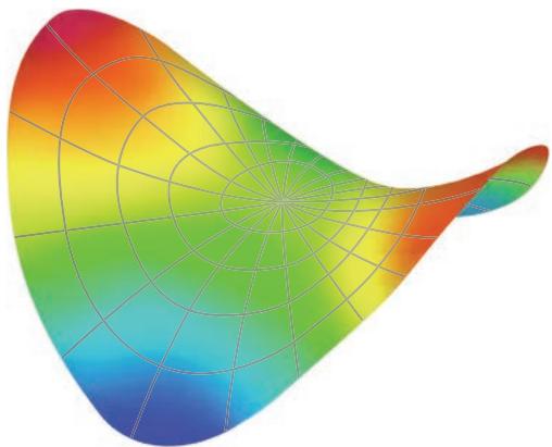
Figure 1-5 Zernike polynomial representation of astigmatism. (Redrawn by Cyndie C. H. Wooley. Origi nal image courtesy of Roger F. Steinert, MD.)
Coma and trefoil
With coma, a third-order aberration, light rays at one edge of the pupil come into focus before those at the opposite edge do. The rays entering the system do not focus on a plane; rather, one edge of the incoming beam focuses either in front of or behind the opposite edge of the beam (see Figure 1-7). The image generated by an incoming light beam pass ing through an optical system with a coma aberration would appear “smeared,” looking somewhat like a comet, with a zone of sharp focus at one edge tailing off to a fuzzy focus at the opposite edge of the beam. Coma is common in patients with decentered corneal grafts, keratoconus, and decentered laser ablations.
Trefoil, also a third-order aberration, can occur after refractive surgery. Trefoil pro duces less degradation in image quality than does coma of similar RMS magnitude (see Figure 1-8).
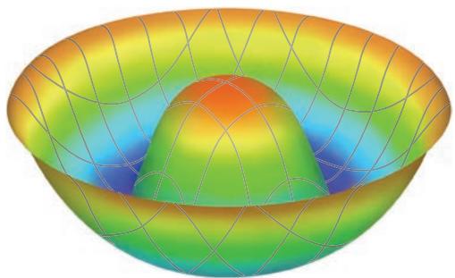
Figure 1-6 Zernike polynomial representation of spherical aberration. (Redrawn by Cyndie C. H. Wooley. Original image courtesy of Roger F. Steinert, MD.)

Figure 1-7 Zernike polynomial representation of coma. (Redrawn by Cyndie C. H. Wooley. Original image courtesy of Roger F. Steinert, MD.)
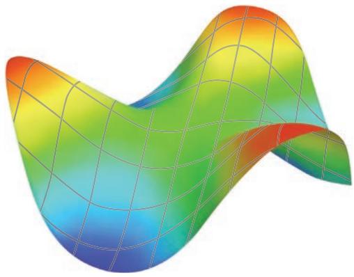

Figure 1-8 Zernike polynomial representation of trefoil. (Redrawn by Cyndie C. H. Wooley. Original image courtesy of Roger F. Steinert, MD.)