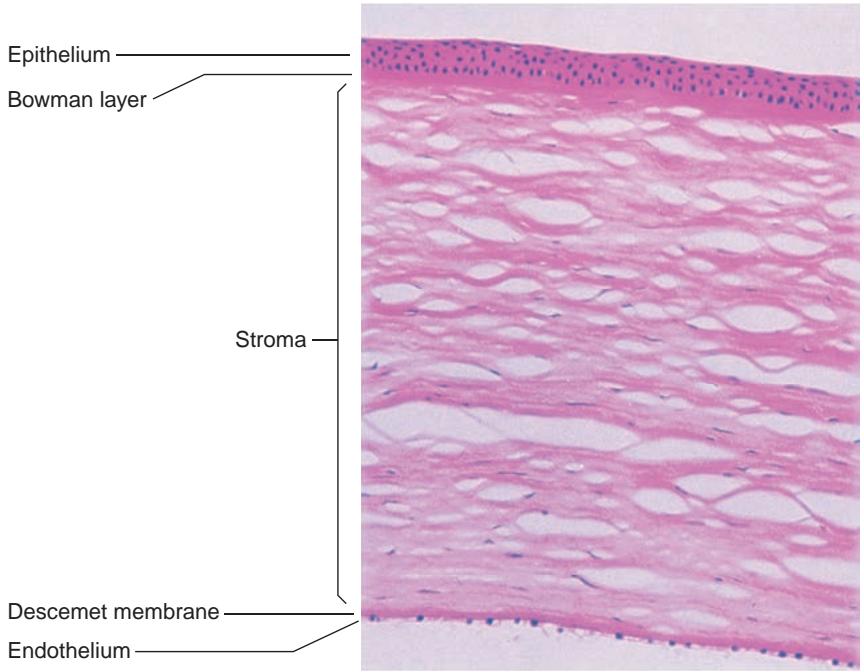

Figure 1-4 The layers of the normal cornea. The epithelium is composed of 4–6 cell layers, but it can increase in thickness to maintain a smooth surface (H&E, x32).
Zones of the Cornea
The cornea is aspheric, although the central portion of the anterior corneal surface has been described as a spherocylindrical convex mirror. The central cornea is typically 3.00 diopters (D) steeper than the periphery, a positive shape factor (prolate).
Clinically, the cornea can be divided into 5 zones (Fig 1-5) as follows:
central zone: 1–3 mm in diameter; closely resembles a spherical surface
paracentral zone: a 3–4 mm “doughnut” surrounding the central zone; has an outer diameter of 7–8 mm and progressively flattens out from the center
apical zone: comprises the paracentral and central zones, used in contact lens fit ting; is primarily responsible for the refractive power of the cornea
peripheral or transitional zone: adjacent to the paracentral zone, has an outer dia meter of approximately 11 mm; is the area of greatest flattening and asphericity in the normal cornea
limbal zone (limbus): where the cornea steepens prior to joining the sclera at the limbal sulcus; outer diameter averages 12 mm

Figure 1-5 Topographic zones of the cornea. (Illustration by Christine Gralapp.)
Other corneal reference definitions include the following:
optical zone: the portion of the cornea that overlies the entrance pupil of the iris
corneal apex: the point of maximum curvature, typically temporal to the center of the pupil
corneal vertex: the point located at the intersection of the line of fixation and the corneal surface; represented by the corneal light reflex when illuminated coaxially with fixation. It is the center of the keratoscopic image and does not necessarily cor respond to the point of maximum curvature at the corneal apex (Fig 1-6)
Corneal Epithelium
The hydrophobic corneal epithelium is composed of 4–6 layers, which include 1–2 layers of superficial squamous cells, 2–3 layers of broad wing cells, and an innermost layer of co lumnar basal cells. It is 40–50 µm thick (see Fig 1-4; also see the Pachymetry section in Chapter 2). The epithelium and tear film form an optically smooth surface. Tight junctions between superficial epithelial cells prevent penetration of tear fluid into the stroma. Con tinuous proliferation of limbal stem cells gives rise to the other layers, which subsequently differentiate into superficial cells. With maturation, these differentiated cells become coated with microvilli on their outermost surface (glycocalyx) and then desquamate into the tears. The process of differentiation takes approximately 7–14 days. Basal epithelial cells secrete a continuous, 50-nm-thick basement membrane, which is composed of type IV collagen, laminin, and other proteins. Corneal clarity depends on the tight packing of epithelial cells, which results in a layer with a nearly uniform refractive index and minimal light scattering.
Bowman Layer
Bowman layer lies anterior to the corneal stroma. Previously considered a membrane, Bowman layer is rather the acellular condensate of irregularly arranged collagen fibrils at the most anterior portion of the stroma (see Fig 1-4). This layer is 15 µm thick and helps maintain the shape of the cornea. If disrupted, it will not regenerate.

Figure 1-6 Corneal vertex and apex. (Illustration by Christine Gralapp.)
Corneal Stroma
The corneal stroma accounts for roughly 90% of total corneal thickness (see Fig 1-4). It is composed of stromal cells (keratocytes), fibers, and an extracellular matrix. The anterior stroma is denser than the posterior stroma due to an increased number of keratocytes and greater interweaving of collagen lamellae. The anterior 40% of the corneal stroma has twice the tensile strength of the posterior 60%. This difference between the anterior and posterior stroma may play a role in the occurrence of corneal ectasia following deep excimer laser ablation.
Keratocytes vary in size and density throughout the stroma and form a 3-dimensional network throughout the cornea. These cells, which are flattened fibroblasts, are located be tween the stromal collagen lamellae (Fig 1-7) and continually digest and manufacture stromal molecules. Keratocyte density declines with age and also following laser refractive surgery.
The extracellular matrix of the corneal stroma is formed from collagens and proteo glycans. Type I and type V fibrillar collagens are intertwined with filaments of type VI collagen. The major corneal proteoglycans are decorin (associated with dermatan sulfate) and lumican (associated with keratan sulfate).
The even distribution of keratocytes, fibers, and the extracellular matrix in the corneal stroma is necessary for a clear cornea. Corneal transparency also depends on maintaining the water content of the corneal stroma at 78%. Corneal hydration is largely controlled by intact epithelial and endothelial barriers and the functioning of the endothelial pump, which is linked to an ion-transport system controlled by temperature-dependent enzymes such as Na+, K+ -ATPase. In addition, negatively charged stromal glycosaminoglycans tend to repel each other, producing a swelling pressure (SP). Because intraocular pressure (IOP) tends to compress the cornea, the overall imbibition pressure of the corneal stroma is calculated as IOP—SP. Corneal hydration varies from anterior to posterior and increases closer to the endothelium.
