CLINICAL PEARL
In patients with visually significant cataract and pterygium, a staged surgical ap proach is indicated. After the pterygium is excised and the corneal contour has stabilized, cataract surgery can be planned; this approach can lead to improved long-term refractive results (Fig 7-5). xvii
xviii • Introduction to Section 8
New and Revised Images
All the images were carefully reviewed, and many new, high-quality images were added to help illustrate clinical points. Existing images were edited to make them more informative, and poor-quality images were removed.
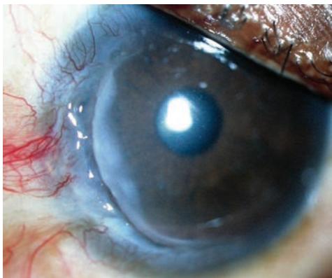
Figure 13-25 Mooren ulcer. Temporal cornea in the right eye demonstrates severe peripheral corneal ulceration. (Courtesy of Arie L. Marcovich, MD, PhD.)
Multimedia
Over 30 new videos were added; many of these videos provide excellent demonstrations of surgical procedures. Three new animations illustrate complex processes to aid viewer understanding.
Online Appendix
This major revision includes access to an online appendix, a microbiology primer that serves as a refresher for readers who may wish to refamiliarize themselves with topics covered in this volume.
Organizational Changes and Readability
Content has been reorganized for a more logical flow and easier readability, with emphasis on teaching the information senior residents need to know. The chapter on therapeutic interventions for ocular surface disorders was moved to follow the 2 chapters on ocular surface disorders. In addition, the coverage of corneal dystrophies and corneal ectasia was split into 2 chapters. Long paragraphs of text have been split into smaller subsections and bulleted lists. Tables that clarify material in the text and present related differential diagnoses in a comparative way have been added.
Many thanks to the BCSC Section 8 committee for their diligent work and commitment to improving an already excellent cornea and external disease text.
Objectives
Upon completion of BCSC Section 8, External Disease and Cornea, the reader should be able to
describe the anatomy of the external eye and cornea
explain the overall strategy, examination, and technology used for systematic evaluation of the cornea and the external eye
identify the distinctive clinical signs and treatment of specific diseases of the ocular surface
identify the most common underlying causes of dry eye and the appropriate approach to management
name the distinguishing clinical features of congenital diseases of the cornea and sclera
identify unique clinical features that help differentiate the more common corneal dystrophies and describe an appropriate management strategy for each
list the risk factors, clinical signs, and breadth of management options of corneal ectasia
describe the basic principles and the clinical approach to the diagnosis and treatment of viral, bacterial, fungal, and parasitic keratitis
identify the common corneal manifestations of systemic disease and describe their treatments
define an approach to the evaluation, diagnosis, and management of immune-related diseases of the external eye and cornea
list the risk factors, diagnosis, and treatment of neoplastic disease of the cornea and the external eye
describe the indications for and techniques of surgical procedures used in the management of corneal disease and trauma
discuss common surgical interventions for ocular surface disorders such as pterygium and corneal melts
explain the role of full-thickness and contemporary partial-thickness keratoplasty in the treatment of corneal disease
Structure and Function of the External Eye and Cornea

This chapter includes a related video. Go to www.aao.org/bcscvideo_section08 or scan the QR code in the text to access this content.

This chapter includes a related activity. Go to www.aao.org/bcscactivity_section08 or scan the QR code in the text to access this content.

Indicates selected key points within the chapter.