Основные положения
Highlights
Внешние структуры глаза обладают как анатомическими, так и иммунологическими механизмами защиты от инфекций и других заболеваний глаз.
The external eye has both anatomical and immunologic defense mechanisms for protection against infection and other ocular conditions.
Знание анатомии роговицы имеет жизненно важное значение для понимания классификаций заболеваний роговицы и освоения развивающихся методов кератопластики.
Knowledge of corneal anatomy is vital for an understanding of corneal disease clas sifications and mastery of evolving keratoplasty techniques.
Роговица представляет собой прозрачную аваскулярную ткань. Прозрачность обусловлена организацией кератоцитов, волокон и внеклеточного матрикса в строме роговицы, а также тонким балансом сил, контролирующих содержание воды в строме.
The cornea is a transparent and avascular tissue. Transparency results from the organization of keratocytes, fibers, and the extracellular matrix within the corneal stroma as well as the delicate balance of forces controlling stromal water content.
Веки
Eyelids
Функции век включают защиту глаза, распределение слезы, очищение поверхности глаза, регулирование светового воздействия и участие в формировании слезной пленки. За исключением тонких пушковых волос, ресницы (cilia) являются единственными волосами века. Ресницы улавливают мелкие частицы и работают как сенсоры для стимуляции рефлекторного закрытия век. Моргание стимулирует работу лакримального насоса для выделения слез, которые затем распределяются по роговице, вымывая инородные материалы. Большинство людей моргают в среднем 10–15 раз в минуту в состоянии покоя, 20 раз в минуту или чаще во время разговора и всего 5 раз в минуту при концентрации (например, при чтении). Частота моргания также меняется при различных положениях взора. Круговая мышца глаза (m. orbicularis oculi), иннервируемая CN VII, закрывает верхнее и нижнее веки (рис. 1-1). Мышца, поднимающая верхнее веко (m. levator palpebrae), иннервируемая CN III, прикрепляется к тарзальной пластинке и коже и поднимает верхнее веко. Мышца Мюллера, иннервируемая симпатическими нервами, также способствует подъему верхнего века. Нижняя тарзальная мышца помогает оттягивать нижнее веко.
The functions of the eyelid include eye protection, tear distribution, ocular surface cleaning, regulation of light exposure, and contribution to the tear film. Except for fine vellus hairs, the eyelashes (cilia) are the only hairs of the eyelid. Eyelashes catch small particles and work as sensors to stimulate reflex eyelid closure. Blinking stimulates the lacrimal pump to release tears, which are then spread across the cornea, flushing foreign material. Most individuals blink an average of 10–15 times per minute at rest, 20 times per minute or more during a conversation, and as few as 5 times per minute when concentrating (eg, reading). Blink frequency also changes in different positions of gaze. The orbicularis oculi muscle, which is innervated by cranial nerve (CN) VII, closes the upper and lower eyelids (Fig 1-1). The leva tor palpebrae muscle, innervated by CN III, inserts into the tarsal plate and skin and elevates the upper eyelid. The Müller muscle, innervated by sympathetic nerves, also contributes to the elevation of the upper eyelid. The inferior tarsal muscle helps retract the lower eyelid.
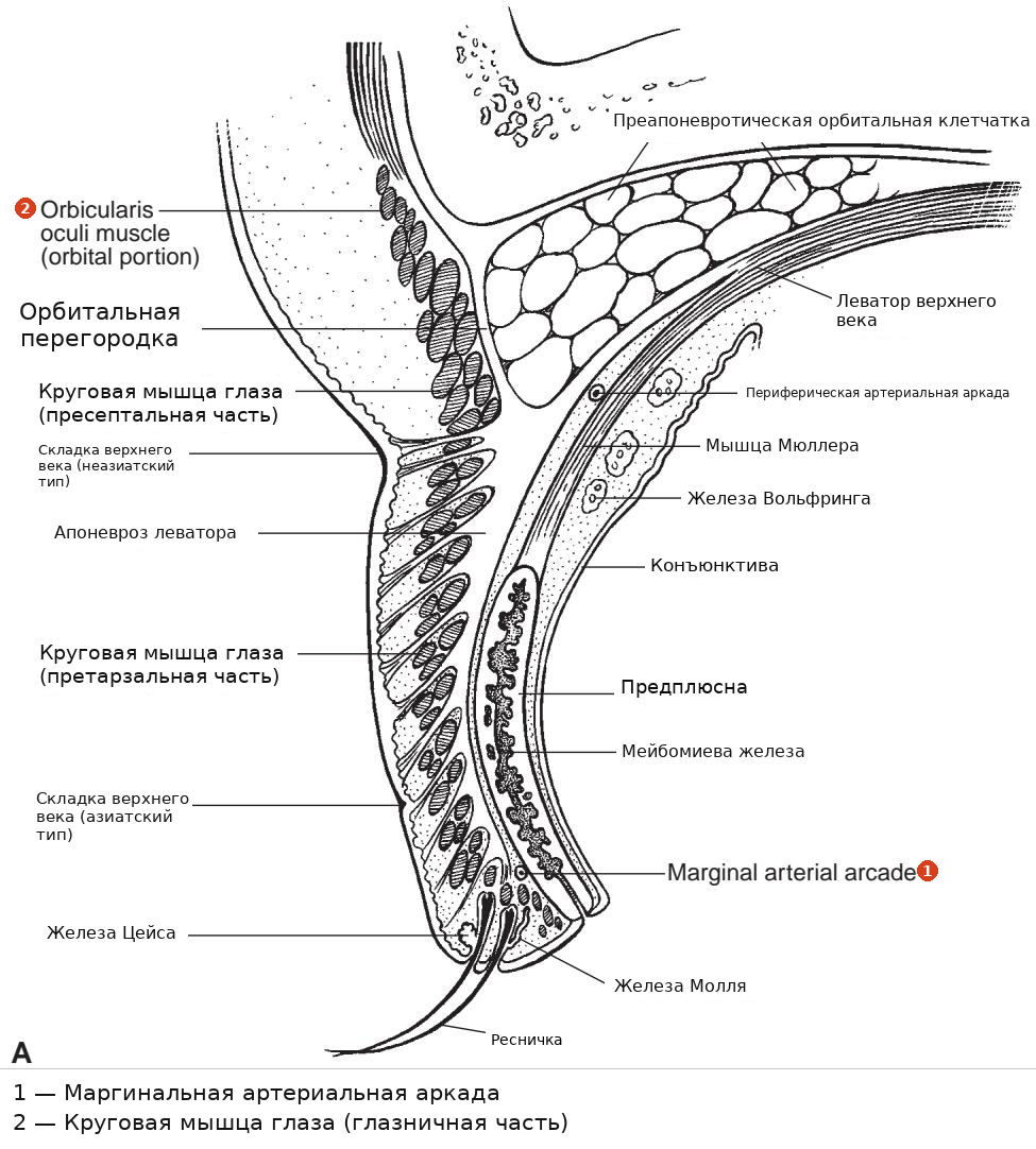
Рисунок 1-1 Веко. A, Иллюстрация поперечного сечения верхнего века.
Figure 1-1 The eyelid. A, Illustration of a cross section of the upper eyelid.
(Продолжение)
(Continued)
Эпидермис век резко меняется с кератинизированного на некератинизированный многослойный плоский эпителий в области слизисто-кожного перехода края века, вдоль ряда устий мейбомиевых желез. В коже века присутствуют голокриновые сальные железы и эккриновые потовые железы. Рядом с краем века расположены апокриновые потовые железы (железы Молля) и многочисленные модифицированные сальные железы (железы Цейса).
The epidermis of the eyelids abruptly changes from keratinized to nonkeratinized stratified squamous epithelium at the mucocutaneous junction of the eyelid margin, along the row of meibomian gland orifices. Holocrine sebaceous glands and eccrine sweat glands are present in the eyelid skin. Near the eyelid margin are apocrine sweat glands (the glands of Moll) and numerous modified sebaceous glands (the glands of Zeis).
Для дополнительного обсуждения анатомии век см. BCSC Раздел 2, Fundamentals and Principles of Ophthalmology, и Раздел 7, Oculofacial Plastic and Orbital Surgery.
For additional discussion of eyelid anatomy, see BCSC Section 2, Fundamentals and Principles of Ophthalmology, and Section 7, Oculofacial Plastic and Orbital Surgery.
Argilés M, Cardona G, Pérez-Cabré E, Rodríguez M. Частота моргания и неполное моргание в шести различных контролируемых условиях чтения на бумажном и электронном носителях. Invest Ophthalmol Vis Sci. 2015;56(11):6679–6685.
Argilés M, Cardona G, Pérez-Cabré E, Rodríguez M. Blink rate and incomplete blinks in six different controlled hard-copy and electronic reading conditions. Invest Ophthalmol Vis Sci. 2015;56(11):6679–6685.
Lin LK, Gokoffski KK. Eyelids and the corneal surface. In: Mannis MJ, Holland EJ, eds. Cornea. Vol 1. 4th ed. Elsevier; 2017:40–45.
Lin LK, Gokoffski KK. Eyelids and the corneal surface. In: Mannis MJ, Holland EJ, eds. Cornea. Vol 1. 4th ed. Elsevier; 2017:40–45.
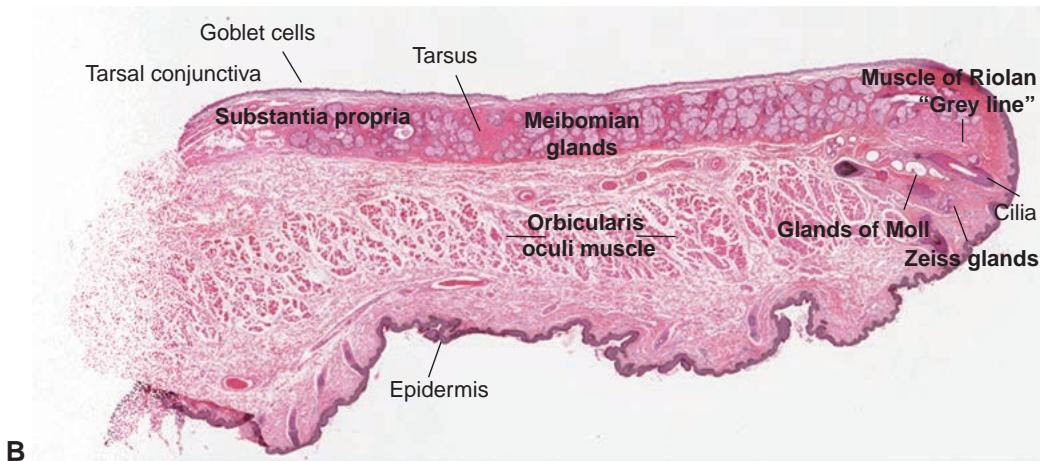

Рисунок 1-1 (продолжение) B, Срез нормального века, окрашенный гематоксилином и эозином (H&E). (Иллюстрация части A выполнена Christine Gralapp, часть B © American Academy of Ophthalmology 2020.)
Figure 1-1 (continued) B, Hematoxylin-eosin (H&E) stained section of the normal eyelid. (Part A illustration by Christine Gralapp, part B © American Academy of Ophthalmology 2020.)
Лакримальный функциональный комплекс
Lacrimal Functional Unit
Лакримальный функциональный комплекс (LFU) включает слезные железы, поверхность глаза и веки, а также сенсорные и моторные нервы, которые соединяют эти компоненты (Видео 1-1; рис. 1-2). LFU отвечает за следующее:
The lacrimal functional unit (LFU) comprises the lacrimal glands, ocular surface, and eyelids, as well as the sensory and motor nerves that connect these components (Video 1-1; Fig 1-2). The LFU is responsible for the following:
регуляцию, выработку, здоровье и целостность слезной пленки (выполнение смазывающей, антимикробной и трофической функций)
regulation, production, health, and integrity of the tear film (carrying out lubricat ing, antimicrobial, and nutritional roles)
здоровье поверхности глаза (поддержание прозрачности роговицы и популяции стволовых клеток поверхности)
health of the ocular surface (maintaining corneal transparency and the surface stem cell population)
качество изображения, проецируемого на сетчатку
quality of the image projected onto the retina

VIDEO 1-1 Восприятие прикосновения и иннервация лакримального функционального комплекса.
VIDEO 1-1 Perception of touch and innervation of the lacrimal functional unit.
Модифицировано с разрешения Pflugfelder SC, Beuerman RW, Stern ME, eds. Dry Eye and Ocular Surface Disorders. Опубликовано CRC Press. © Marcel Dekker; 2004, воспроизведено по договоренности с Taylor & Francis Books UK.
Modified with permission from Pflugfelder SC, Beuerman RW, Stern ME, eds. Dry Eye and Ocular Surface Disorders. Published by CRC Press. © Marcel Dekker; 2004, reproduced by arrangement with Taylor & Francis Books UK.

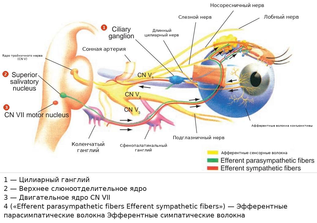
Рисунок 1-2 Сенсорные и моторные нервы, соединяющие компоненты лакримального функционального комплекса. CN = черепной нерв. (Модифицировано с разрешения Pflugfelder SC, Beuerman RW, Stern ME, eds. Dry Eye and Ocular Surface Disorders. Опубликовано CRC Press. @ Marcel Dekker; 2004, воспроизведено по договоренности с Taylor & Francis Books UK.)
Figure 1-2 The sensory and motor nerves connecting the components of the lacrimal func tional unit. CN = cranial nerve. (Modified with permission from Pflugfelder SC, Beuerman RW, Stern ME, eds. Dry Eye and Ocular Surface Disorders. Published by CRC Press. @ Marcel Dekker; 2004, reproduced by arrangement with Taylor & Francis Books UK.)
LFU реагирует на воздействие факторов окружающей среды, эндокринологические и кортикальные влияния. Афферентный компонент LFU опосредован ноцицепторами поверхности глаза и тройничного нерва, которые образуют синапсы с эфферентными нервами (автономными и моторными нервами) в стволе головного мозга. Автономные нервные волокна иннервируют мейбомиевы железы, бокаловидные клетки конъюнктивы и слезные железы. Моторные нервные волокна иннервируют круговую мышцу для инициации моргания. Во время моргания мейбомиевы железы выделяют липиды, и слеза восполняется из нижнего слезного мениска и распределяется по роговице, в то время как избыток слезы направляется в слезные точки.
The LFU responds to environmental, endocrinologic, and cortical influences. The afferent component of the LFU is mediated through ocular surface and trigeminal no ciceptors, which synapse with the efferent nerves (autonomic and motor nerves) in the brainstem. The autonomic nerve fibers innervate the meibomian glands, conjunctival gob let cells, and lacrimal glands. The motor nerve fibers innervate the orbicularis muscle to initiate blinking. During blinking, the meibomian glands express lipid, and the tears are replenished from the inferior tear meniscus and spread across the cornea while excess tears are directed into the lacrimal puncta.