Pflugfelder SC, Beuerman RW, Stern ME, eds. Dry Eye and Ocular Surface Disorders. CRC Press/Taylor & Francis; 2004.
Pflugfelder SC, Beuerman RW, Stern ME, eds. Dry Eye and Ocular Surface Disorders. CRC Press/Taylor & Francis; 2004.
Слезная пленка
Tear Film
В настоящее время считается, что слезная пленка представляет собой смешанный гель, состоящий из растворимой слизи, жидкостей и белков, которые вырабатываются слезной железой, бокаловидными клетками конъюнктивы и эпителием поверхности (рис. 1-3). Этот гидрофильный гель перемещается по гликокаликсу поверхностных эпителиальных клеток роговицы и покрывается липидным слоем, вырабатываемым мейбомиевыми железами.
The tear film is currently thought to be a mixed gel consisting of soluble mucus, fluids, and proteins that are contributed by the lacrimal gland, conjunctival goblet cells, and surface epithelium (Fig 1-3). This hydrophilic gel moves over the glycocalyx of the superficial corneal epithelial cells and is topped by a lipid layer produced by the meibomian glands.
Здоровая слезная пленка имеет решающее значение как для хорошего зрения, так и для здоровья глаза. Функции слезной пленки включают
A healthy tear film is critical for both good vision and a healthy eye. Functions of the tear film include
поддержание гладкой оптической поверхности между морганиями
maintaining a smooth optical surface between blinks
вклад в рефракционную силу глаза через границу раздела воздух–слезная пленка
contributing to refractive power of the eye through the air–tear film interface
удаление раздражителей, патогенов, токсинов, аллергенов и детрита
removing irritants, pathogens, toxins, allergens, and debris
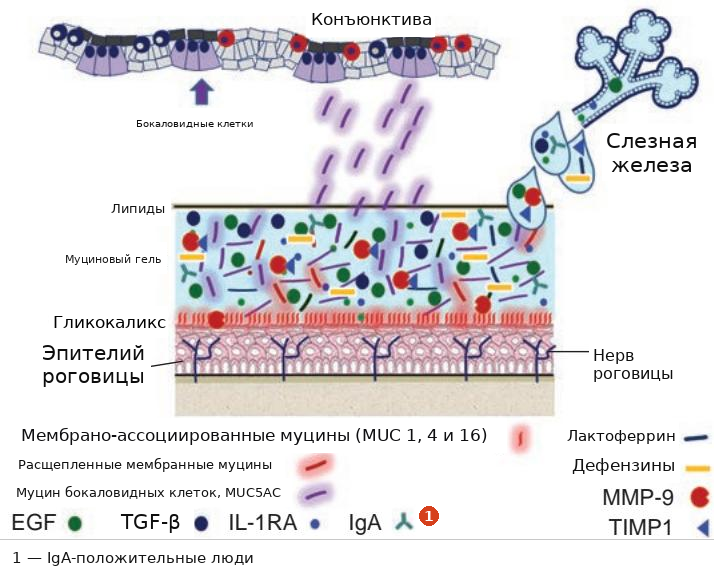
Рисунок 1-3 Слезная пленка состоит из смешанного муцинового/водного слоя, вырабатываемого слезными железами, бокаловидными клетками конъюнктивы и эпителием поверхности. Сверху она покрыта липидным слоем, вырабатываемым мейбомиевыми железами. Ее функции включают смазку (муцины), заживление (эпидермальный фактор роста [EGF]) и защиту роговицы от инфекции (лактоферрин, дефензины, иммуноглобулин A [IgA]). При воспалении слезной пленки она вырабатывает антагонист рецептора интерлейкина 1 (IL-1RA), трансформирующий ростовой фактор β (TGF-β) и тканевый ингибитор матриксной металлопротеиназы 1 (TIMP 1). MMP-9 = матриксная металлопротеиназа 9. (Модифицировано с разрешения Pflugfelder SC. Tear dysfunction and the cornea: LXVIII Edward Jackson Memorial Lecture. Am J Ophthalmol. 2011;152(6):902, с разрешения Elsevier.)
Figure 1-3 The tear film consists of a mixed mucin/aqueous layer produced by the lacrimal glands, conjunctival goblet cells, and surface epithelium. It is topped by a lipid layer produced by the meibomian glands. Its functions include lubrication (mucins), healing (epidermal growth factor [EGF]), and protection of the cornea against infection (lactoferrin, defensins, immunoglobu lin A [IgA]). When the tear film is inflamed, it produces interleukin 1 receptor antagonist (IL-1RA), transforming growth factor $ \beta $ (TGF-$ \beta $), and tissue inhibitor of matrix metalloproteinase 1 (TIMP 1). MMP-9 = matrix metalloproteinase 9. (Modified with permission from Pflugfelder SC. Tear dysfunction and the cornea: LXVIII Edward Jackson Memorial Lecture. Am J Ophthalmol. 2011;152(6):902, with permission from Elsevier.)
облегчение диффузии кислорода и других питательных веществ к роговице и конъюнктиве
facilitating the diffusion of oxygen and other nutrients to the cornea and conjunctiva
поддержание гомеостаза нормальной глазной флоры
maintaining homeostasis of the normal ocular flora
вклад в антимикробную защиту поверхности глаза
contributing to the antimicrobial defense of the ocular surface
Таким образом, поддержание слезной пленки имеет решающее значение для нормального функционирования роговицы.
Maintenance of the tear film is thus critical to normal corneal function.
Пфлугфельдер SC. Дисфункция слезной пленки и роговица: LXVIII Мемориальная лекция имени Эдварда Джексона.
Pflugfelder SC. Tear dysfunction and the cornea: LXVIII Edward Jackson Memorial Lecture.
Am J Ophthalmol. 2011;152(6):900–909.
Am J Ophthalmol. 2011;152(6):900–909.
Конъюнктива
Conjunctiva
Конъюнктиву можно условно разделить на 3 географические зоны следующим образом:
The conjunctiva can be broadly divided into 3 geographic zones as follows:
вековая, или тарзальная: начинается у слизисто-кожного перехода верхнего и нижнего век и покрывает внутреннюю поверхность века; она плотно сращена с подлежащим тарзусом и прикрепляется к лимбу
palpebral, or tarsal: starts at the mucocutaneous junction of the upper and lower eyelid and covers the inner eyelid; it is tightly adherent to the underlying tarsus and inserts into the limbus
бульбарная: покрывает поверхность глазного яблока и рыхло прикреплена к капсуле Тенона; прикрепляется к лимбу
bulbar: covers the ocular surface and is loosely attached to the Tenon capsule; it inserts into the limbus
сводчатая: покрывает верхний и нижний своды
forniceal: covers the superior and inferior fornices
Полулунная складка (plica semilunaris) представляет собой серповидную вертикальную складку конъюнктивы, расположенную в медиальном углу глаза. Каранкула — мясистое овальное образование высотой примерно 5 мм и шириной 3 мм — прикреплена к нижне-медиальной стороне полулунной складки и содержит бокаловидные клетки и слезную ткань, а также волоски, сальные и потовые железы. В этой области также находится lacus lacrimalis (слезное озеро) — небольшое треугольное пространство, где скапливаются слезы после омывания поверхности глазного яблока.
The plica semilunaris is a crescent-shaped vertical fold of conjunctiva, located at the medial angle of the eye. The caruncle—a fleshy, ovoid mass approximately 5 mm high and 3 mm wide—is attached to the inferomedial side of the plica semilunaris and contains goblet cells and lacrimal tissue, as well as hairs, sebaceous glands, and sweat glands. This area also contains the lacus lacrimalis (lacrimal lake), a small triangular space where tears accumulate after bathing the ocular surface.
Морфология клеток эпителия конъюнктивы варьирует от многорядного кубического над тарзусом и цилиндрического в сводах до плоского на глазном яблоке. Бокаловидные клетки составляют до 10% базальных клеток эпителия конъюнктивы и наиболее многочисленны в вековой конъюнктиве, нижне-назальной бульбарной конъюнктиве и в области полулунной складки.
The cell morphology of the conjunctival epithelium varies from stratified cuboidal over the tarsus and columnar in the fornices to squamous on the globe. Goblet cells account for up to 10% of basal cells of the conjunctival epithelium and are most numerous in the palpe bral conjunctiva, the inferonasal bulbar conjunctiva, and the area of the plica semilunaris.
Собственная пластинка (substantia propria) конъюнктивы состоит из рыхлой соединительной ткани. Присутствует конъюнктивально-ассоциированная лимфоидная ткань (CALT), состоящая из лимфоцитов и других лейкоцитов, особенно в сводах. Лимфоциты взаимодействуют с клетками слизистого эпителия посредством взаимных регуляторных сигналов, опосредованных факторами роста, цитокинами и нейропептидами.
The substantia propria of the conjunctiva consists of loose connective tissue. Conjunctiva- associated lymphoid tissue (CALT), which consists of lymphocytes and other leukocytes, is present, especially in the fornices. Lymphocytes interact with mucosal epithelial cells through reciprocal regulatory signals mediated by growth factors, cytokines, and neuropeptides.
Кровоснабжение вековой конъюнктивы осуществляется совместно с веками. Бульбарная конъюнктива снабжается передними цилиарными артериями, которые отходят от мышечных ветвей глазной артерии. Эти капилляры имеют фенестры и пропускают жидкость, что вызывает хемоз (отек конъюнктивы) в ответ на аллергические или другие воспалительные процессы.
The palpebral conjunctiva shares its blood supply with the eyelids. The bulbar con junctiva is supplied by the anterior ciliary arteries, which arise from muscular branches of the ophthalmic artery. These capillaries are fenestrated and leak fluid, producing chemo- sis (conjunctival swelling), as a response to allergies or other inflammatory events.
Склера
Sclera
Склера представляет собой непрозрачную белую фиброзную ткань, которая простирается от лимба роговицы до зрительного нерва, где она сливается с ним, образуя твердую оболочку зрительного нерва. Она разделена на 3 слоя (от наружного к внутреннему): эписклера, строма и lamina fusca. Строма склеры состоит из коллагеновых волокон, которые имеют различную ориентацию, степень разделения и диаметр, что обуславливает непрозрачный вид склеры в отличие от роговицы. Кровоснабжение склеры осуществляется передними и задними цилиарными артериями, а отток идет через вортикозные вены. Толщина склеры варьирует от 0.3 мм до 1 мм; она наиболее тонкая за местами прикрепления прямых мышц. Иннервируется цилиарными нервами черепного нерва V1.
The sclera is the opaque, white, fibrous tissue that extends from the corneal limbus to the optic nerve, where it merges to form the dural sheath of the optic nerve. It is divided into 3 layers (from outermost inward): episclera, stroma, and lamina fusca. The scleral stroma is composed of collagen fibers, which have varied orientation, separation, and diameter, resulting in the sclera’s opaque appearance in contrast to the cornea. The sclera receives its vascular supply from the anterior and posterior ciliary arteries and drains through the vortex veins. The thickness of the sclera ranges from 0.3 mm to 1 mm; it is thinnest behind the insertions of the rectus muscles. It is innervated by the ciliary nerves of cranial nerve V1.
Для дополнительного обсуждения склеры см. BCSC Раздел 2, Fundamentals and Principles of Ophthalmology, и Раздел 4, Ophthalmic Pathology and Intraocular Tumors.
For additional discussion of the sclera, see BCSC Section 2, Fundamentals and Princi ples of Ophthalmology, and Section 4, Ophthalmic Pathology and Intraocular Tumors.
Роговица
Cornea
Роговица представляет собой прозрачную аваскулярную ткань, состоящую из 5 слоев (Задание 1-1; Рис 1-4): эпителия, слоя Боумена, стромы, мембраны Десцеме и эндотелия; они обсуждаются в следующих подразделах.
The cornea is a transparent, avascular tissue that consists of 5 layers (Activity 1-1; Fig 1-4): epithelium, Bowman layer, stroma, Descemet membrane, and endothelium; these are dis cussed in the subsections that follow.
У взрослых размер роговицы составляет примерно 11–12 мм по горизонтали и 10–11 мм по вертикали. Ее толщина в центре составляет 500–600 мкм, постепенно увеличиваясь к периферии.
In adults, the cornea measures approximately 11–12 mm horizontally and 10–11 mm vertically. It is 500–600 µm thick at its center, gradually increasing in thickness toward the periphery.

ЗАДАНИЕ 1-1 Слои роговицы и соответствующие конфокальные изображения.
ACTIVITY 1-1 Corneal layers and corresponding confocal images.
Конфокальные изображения предоставлены Danielle Trief, MD, и David D. Verdier, MD.
Confocal images courtesy of Danielle Trief, MD, and David D. Verdier, MD.


Питание роговицы зависит от диффузии глюкозы из водянистого юмора и диффузии кислорода через слезную пленку. Периферическая часть роговицы снабжается кислородом из лимбального кровотока. Плотность нервных окончаний в роговице является одной из самых высоких в организме, а чувствительность роговицы в 100 раз превышает чувствительность конъюнктивы. Сенсорные нервные волокна отходят от длинных цилиарных нервов, проникая в роговицу в глубокой периферической строме около лимба и направляясь вперед, формируя субэпителиальное сплетение.
For nutrition, the cornea depends on diffusion of glucose from the aqueous humor and diffusion of oxygen through the tear film. The peripheral cornea is supplied with oxygen from the limbal circulation. The density of nerve endings in the cornea is among the high est in the body, and the sensitivity of the cornea is 100 times that of the conjunctiva. Sen sory nerve fibers extend from the long ciliary nerves, penetrating the cornea in the deep peripheral stroma near the limbus and coursing anteriorly to form a subepithelial plexus.